

Updates on Geographical Dispersion of Leishmania Parasites Causing Cutaneous Affections in Algeria
- PMID: 33669099
- PMCID: PMC7996526
- DOI: 10.3390/pathogens10030267
Updates on Geographical Dispersion of Leishmania Parasites Causing Cutaneous Affections in Algeria
Abstract
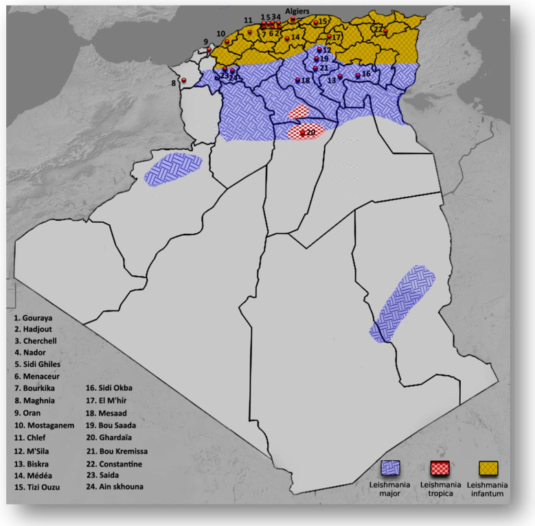
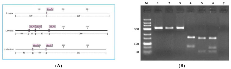
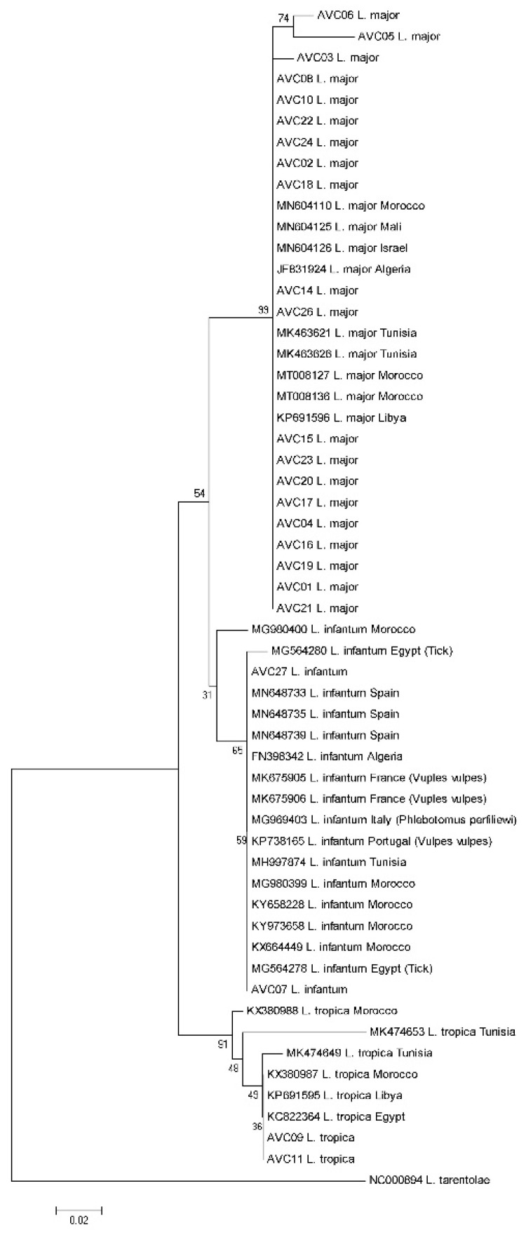
Leishmaniases are neglected tropical diseases of public health concern in Algeria. To update the geographical distribution of Leishmania spp. causing cutaneous affection, we examined a set of Giemsa-stained smears prepared from skin lesions of the patients suspected to have cutaneous leishmaniasis (CL) in various geographical areas in Algeria. The identification of Leishmania parasites was performed using microscopy, conventional PCR, and PCR-RFLP (PCR-Restriction Fragment Length Polymorphism) targeting ITS1-rDNA. Among 32 smears provided from 27 suspected patients with cutaneous lesions, no trace of parasites was observed in the smear of three patients using microscopy and molecular approaches. Furthermore, four patients presented at least two lesions. PCR-RFLP confirmed the presence of Leishmania in 29 smears prepared from 24 patients. Two biopsies, negative after microscopic examination, were found positive by PCR. Of these 29 PCR positive smears (24 patients), 20 were identified using RFLP-PCR as L. major, two as L. tropica, and two as L. infantum. We found L. major infected patients from Ain skhouna, Biskra, El M'hir, Ghardaïa, M'Sila, and Saida, in agreement with previously reported cases. Furthermore, we highlighted for the first time, the identification of L. major in the patients from Bourkika, Bou Kremissa, Bou Saada Clef, Hajout, Maghnia, Médéa, Menaceur, Messad, Mostaghanem, Nador, Oran, and Sidi Okba. A phylogenetic reconstruction performed with sequences collected from the PCR products confirmed these identifications. Our data provide additional information on the geographical extension of CL caused by L. tropica and L. infantum in Algeria.
Keywords: Leishmania infantum; Leishmania major; Leishmania tropica; PCR–RFLP; cutaneous leishmaniasis.
Conflict of interest statement
The authors declare no conflict of interest.
Figures
References
-
- World Health Organization [(accessed on 30 May 2018)];2018 Available online: https://www.who.int/health-topics/leishmaniasis#tab=tab_1.
-
- Kholoud K., Bounoua L., Sereno D., El Hidan M., Messouli M. Emerging and Re-Emerging Leishmaniases in the Mediterranean Area: What Can Be Learned from a Retrospective Review Analysis of the Situation in Morocco during 1990 to 2010? Microorganisms. 2020;8:1511. doi: 10.3390/microorganisms8101511. - DOI - PMC - PubMed
LinkOut - more resources
Full Text Sources
Other Literature Sources
Miscellaneous
