. 2021 Feb 16;22(4):1961.
doi: 10.3390/ijms22041961.

Ym155 Induces Oxidative Stress-Mediated DNA Damage and Cell Cycle Arrest, and Causes Programmed Cell Death in Anaplastic Thyroid Cancer Cells

Affiliations

Ym155 Induces Oxidative Stress-Mediated DNA Damage and Cell Cycle Arrest, and Causes Programmed Cell Death in Anaplastic Thyroid Cancer Cells

Qinqin Xu et al. Int J Mol Sci. .

Abstract

Anaplastic thyroid cancer (ATC) is one of the most lethal malignancies with a median survival time of about 4 months. Currently, there is no effective treatment, and the development of new therapies is an important and urgent issue for ATC patients. YM155 is a small molecule that was identified as the top candidate in a high-throughput screen of small molecule inhibitors performed against a panel of ATC cell lines by the National Cancer Institute. However, there were no follow-up studies investigating YM155 in ATC. Here, we determined the effects of YM155 on ATC and human primary benign thyroid cell (PBTC) survival with alamarBlue assay. Our data show that YM155 inhibited proliferation of ATC cell lines while sparing normal thyroid cells, suggesting a high therapeutic window. YM155-induced DNA damage was detected by measuring phosphorylation of γ-H2AX as a marker for DNA double-strand breaks. The formamidopyrimidine-DNA glycosylase (FPG)-modified alkaline comet assay in conjunction with reactive oxygen species (ROS) assay and glutathione (GSH)/glutathione (GSSG) assay suggests that YM155-mediated oxidative stress contributes to DNA damage. In addition, we provide evidence that YM155 causes cell cycle arrest in S phase and in the G2/M transition and causes apoptosis, as seen with flow cytometry. In this study, we show for the first time the multiple effects of YM155 in ATC cells, furthering a potential therapeutic approach for ATC.

Keywords: DNA damage; YM155; anaplastic thyroid cancer; apoptosis; cell cycle arrest.


Conflict of interest statement

Paul M. Weinberger is chief executive officer for SpheroFill LLC, which has no competing interest or relationship with the current research. The authors declare no conflict of interest.

Figures

Figure 1
Figure 1
YM155 inhibited proliferation of anaplastic thyroid cancer (ATC) cell lines while sparing normal thyroid cells. ATC cell lines (AD) and primary benign thyroid cells (PBTCs) (E) were treated with varying concentrations of YM155 and incubated for 0, 1, 2, or 3 days. Cell viability was measured using the alamarBlue (Bio-Rad, Oxford, UK) assay. ATC cell lines ACT1 (A) and THJ16T (C) were fast growing and showed dramatic responses to YM155 treatment, even at low doses. ATC cell lines THJ11T (B) and THJ29T (D) demonstrated slower growth, comparatively, and were less responsive to YM155 treatment, with complete proliferation inhibition occurring only at higher doses in THJ29T cells and incomplete proliferation inhibition in THJ11T. (E) Primary benign thyroid cells exhibited slow growth and were unaffected by YM155 at all concentrations and times. IC50 for each cell line: ACT1 = 3.24 nM, THJ16T = 5.102 nM, THJ11T = 73.387 nM, THJ29T =18.6433 nM.
Figure 2
Figure 2
YM155 induced DNA damage in ATC cells while human primary benign thyroid cells were unaffected. ATC cells ACT1, THJ16T, and THJ29T showed increases 4hosphorpho-histone H2AX (γ-H2AX), a DNA repair factor and marker for double-strand DNA breaks, after treatment with 10 nM YM155 for 24 h. Cell line THJ11T exhibited elevated levels of γ-H2AX at baseline, which were not significantly increased by YM155 treatment. PBTCs, which exhibited DNA damage when treated with the positive control bleomycin, showed no evidence of DNA damage with YM155 treatment. (A) Examples of pictures captured by fluorescent microscopy. (B) Foci were counted using JQuantPlus [15,16]. γ-H2AX, a count level data, was analyzed using negative binomial regression and reported with mean and 95% confidence interval. Corresponding p-value is associated with the regression. **** indicates p < 0.0001.
Figure 3
Figure 3
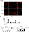
YM155 increased oxidative stress in THJ16T and ACT1. (A) Representative comet assay images. Alkaline comet assay was used to measure single-strand DNA breaks after treatment with 10 nM YM155 for 1 h with or without formamidopyrimidine-DNA glycosylase (Fpg) treatment. Tail moment increased significantly in Fpg+ ACT1 and THJ16T cells, with lower tail moment in Fpg- samples, suggesting that YM155 induces oxidative DNA damage in 1 h. (B) Quantification of tail moment with OpenComet plugin for ImageJ. (C) Effect of YM155 in reactive oxygen species (ROS) assay. Oxidation of H2DCF by intracellular ROS yielded a highly fluorescent product, H2DCFDA, which was detected by microplate reader (Ex/Em 495/529 nm). ROS increased in ATC cells after 1-h treatment with 100 nM YM155. (D) Effect of YM155 in glutathione fluorometric assay. The OPA probe (o-phthalaldehyde) reacted with glutathione (GSH), generating fluorescence at Ex/Em 340/420 nm. To measure glutathione (GSSG), we added a GSH quencher to remove GSH, preventing reaction with OPA, and a reducing agent was then added to remove excess quencher and convert GSSG to GSH. Thus, GSSG can be specifically quantified. Concentration of GSH and GSSG was calculated on the basis of a GSH standard curve. Results were normalized to the control group. **** indicates p < 0.0001; *** indicates p < 0.001; ** indicates p < 0.01.
Figure 4
Figure 4
YM155 induced cell cycle arrested in ATC cells. ATC cell lines ACT1 (A), THJ11T (B), THJ16T (C), and THJ29T (D) treated with YM155 (10 nM), mimosine (360 µM), or control (1/1000 DMSO) for 24 h were fixed and processed, stained with propidium iodide (PI), and analyzed with flow cytometry. Cell cycle analysis calculated the proportion of cells in G0/G1, S, G2, and sub G0. Cells were arrested at S phase and G2/M with YM155 treatment, while the positive control mimosine caused stalling in G0/G1 phase, suggesting that YM155-induced DNA damage occurs during DNA replication.
Figure 5
Figure 5
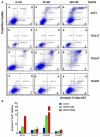
YM155 induced apoptosis in ATC cell lines THJ16T and ACT1. (A) Selected profiles of apoptosis analysis with Alexa Fluo 647 annexin V in ATC cells. The four quadrants 1, 2, 3, and 4 represent live cell, early apoptosis, late apoptosis, and necrosis, respectively. (B) The bar graph indicates the percent of apoptotic and necrotic cells in each group. Annexin V+ indicates annexin V-positive cells. PI+ indicates propidium iodide-positive cells.

References

    1. Saini S., Tulla K., Maker A.V., Burman K.D., Prabhakar B.S. Therapeutic advances in anaplastic thyroid cancer: A current perspective. Mol. Cancer. 2018;17:154. doi: 10.1186/s12943-018-0903-0. - DOI - PMC - PubMed
    1. Neff R.L., Farrar W.B., Kloos R.T., Burman K.D. Anaplastic thyroid cancer. Endocrinol. Metab. Clin. N. Am. 2008;37:525–538. doi: 10.1016/j.ecl.2008.02.003. xi. - DOI - PubMed
    1. O’Neill J.P., Shaha A.R. Anaplastic thyroid cancer. Oral Oncol. 2013;49:702–706. doi: 10.1016/j.oraloncology.2013.03.440. - DOI - PubMed
    1. Perrier N.D., Brierley J.D., Tuttle R.M. Differentiated and anaplastic thyroid carcinoma: Major changes in the American Joint Committee on Cancer eighth edition cancer staging manual. CA Cancer J. Clin. 2018;68:55–63. doi: 10.3322/caac.21439. - DOI - PMC - PubMed
    1. Lin B., Ma H., Ma M., Zhang Z., Sun Z., Hsieh I.Y., Okenwa O., Guan H., Li J., Lv W. The incidence and survival analysis for anaplastic thyroid cancer: A SEER database analysis. Am. J. Transl. Res. 2019;11:5888–5896. - PMC - PubMed

MeSH terms