. 2021 Feb 19;13(4):877.
doi: 10.3390/cancers13040877.

Exosomal HMGB1 Promoted Cancer Malignancy

Affiliations

Exosomal HMGB1 Promoted Cancer Malignancy

Jiaan-Der Wang et al. Cancers (Basel). .

Abstract

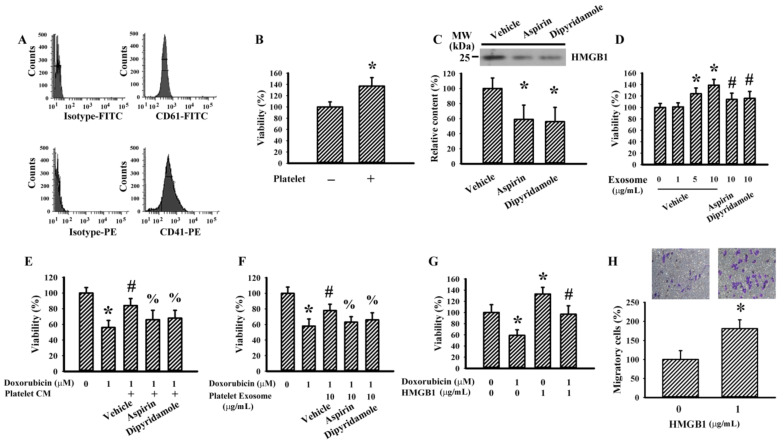
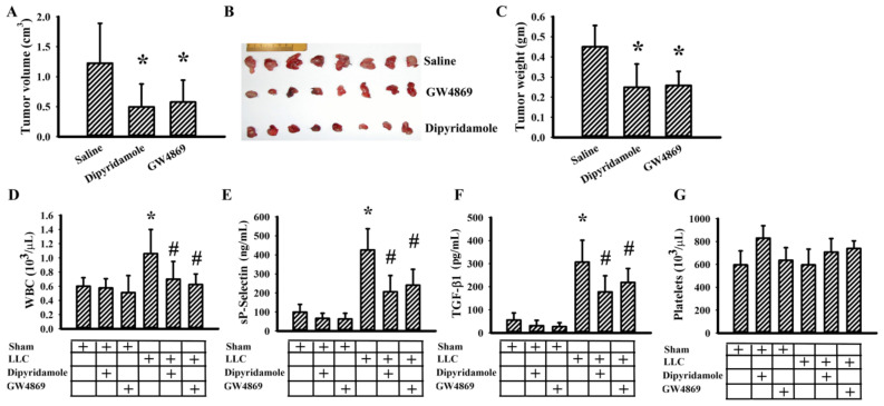
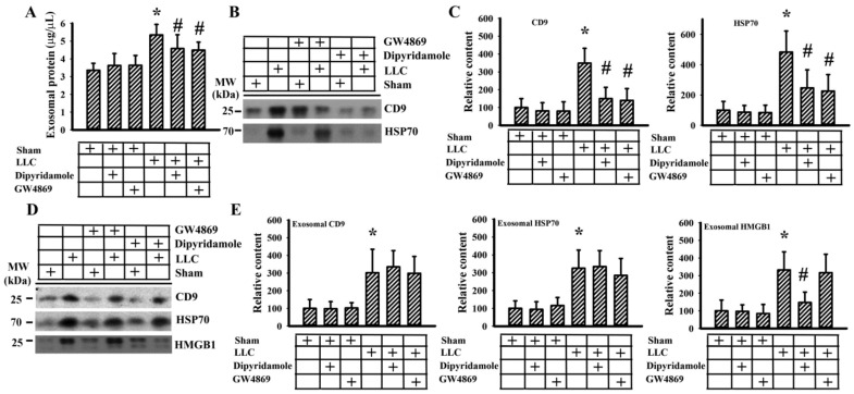
Reciprocal crosstalk between platelets and malignancies underscores the potential of antiplatelet therapy in cancer treatment. In this study, we found that human chronic myeloid leukemia K562 cell-differentiated megakaryocytes and murine platelets produced bioactive substances and these are released into the extracellular space, partly in their exosomal form. High-mobility group box 1 (HMGB1) is a type of exosomal cargo, and the antiplatelet drugs aspirin and dipyridamole interfered with its incorporation into the exosomes. Those released substances and exosomes, along with exogenous HMGB1, promoted cancer cell survival and protected cells from doxorubicin cytotoxicity. In a tumor-bearing model established using murine Lewis lung carcinoma (LLC) cells and C57BL/6 mice, the tumor suppressive effect of dipyridamole correlated well with decreased circulating white blood cells, soluble P-selectin, TGF-β1 (Transforming Growth Factor-β1), exosomes, and exosomal HMGB1, as well as tumor platelet infiltration. Exosome release inhibitor GW4869 exhibited suppressive effects as well. The suppressive effect of dipyridamole on cancer cell survival was paralleled by a reduction of HMGB1/receptor for advanced glycation end-products axis, and proliferation- and migration-related β-catenin, Yes-associated protein 1, Runt-related transcription factor 2, and TGF- β1/Smad signals. Therefore, exosomes and exosomal HMGB1 appear to have roles in platelet-driven cancer malignancy and represent targets of antiplatelet drugs in anticancer treatment.

Keywords: HMGB1; antiplatelet; exosome; malignancy.


Conflict of interest statement

The authors have no conflict of interest to declare.

Figures

Figure 1
Figure 1
Conditioned medium of megakaryocytes promoted T24 cell survival. K562 cells were treated with various concentrations of phorbol 12-myristate 13-acetate (PMA) (0–10 nM) for 4 days. (A) Proteins were extracted and subjected to Western blot analysis with indicated antibodies. Representative blots of four independent experiments are shown. The Western blots have been shown in Figure S1. (B) Cells were harvested and subjected to flowcytometric analysis for the measurement of surface presentation of CD41. (C) Cells were pelleted by centrifugation, spread onto glass slides, and stained with Wright–Giemsa dye. Scale bar: 300 µm. K562 cells were treated with PMA (0 and 10 nM) in the absence or presence of dipyridamole (10 µM), aspirin (100 µg/mL), or cytochalasin D (1 µg/mL) for 4 days. (D) Cells were harvested and subjected to flowcytometric analysis for the measurement of surface presentation of CD41. Bar graphs show relative genomic mean intensity of CD41-PE fluorescence. (E) The cultured media were harvested and centrifuged, and the resultant supernatants were named conditioned medium (CM). Equal amounts of CM and fresh medium were mixed and added to T24 cells (96-well) for 48 h. Cell viability was measured by the 3-(4,5-dimethylthiazol-2-yl)-5-(3-carboxymethoxyphenyl)-2-(4-sulfophenyl)-2H-tetrazolium (MTS) reduction assay. (F) Equal amounts of CM obtained from PMA treatments (0 and 10 nM) and fresh medium were mixed and added to T24 cells (96-well) in the absence or presence of cytochalasin D (1 µg/mL) for 48 h. Cell viability was measured by the MTS reduction assay. * p < 0.05 vs. untreated group and # p < 0.05 vs. PMA (10 nM) group or K562/PMA (10 nM) CM group, n = 4.
Figure 2
Figure 2
Megakaryocytes produced pro-survival exosomes. K562 cells were treated with PMA (10 nM) in the absence or presence of dipyridamole (10 µM) or aspirin (100 µg/mL) for 4 days. Equal amounts of cultured media were subjected to exosome isolation. (A,B) The obtained exosomes were subjected to flowcytometric analysis for the measurement of CD9 positive particles. (C) The obtained exosomes were subjected to the measurement of protein content. (D) The obtained exosomes were subjected to Western blot analysis with indicated antibodies. Representative blots of four independent experiments are shown. (E) Various concentrations of exosomes based on protein contents were added to T24 cells (96-well) for 48 h. Cell viability was measured by the MTS reduction assay. T24 cells were treated with doxorubicin (0 and 2 µM) in the absence or presence of the obtained exosomes (5 µg/mL), dipyridamole (10 µM), or aspirin (100 µg/mL) for 48 h. (F) Cells were harvested and subjected to flowcytometric analysis for the measurement of SubG1 population. (G) Proteins were extracted and subjected to enzymatic assay for the measurement of caspase 3 activity. T24 cells were treated with doxorubicin (0 and 2 µM) in the absence or presence of high-mobility group box 1 (HMGB1) (5 µg/mL) for 48 h. (H) Cells were harvested and subjected to flowcytometric analysis for the measurement of SubG1 population. (I) Proteins were extracted and subjected to enzymatic assay for the measurement of caspase 3 activity. * p < 0.05 vs. untreated group, # p < 0.05 vs. exosome (vehicle, 5 µg/mL) or doxorubicin (2 µM), and % p < 0.05 vs. doxorubicin (2 µM)/exosome (vehicle, 5 µg/mL), n = 4.
Figure 3
Figure 3
Conditioned medium and exosomes of platelets promoted Lewis lung carcinoma (LLC) cell survival. Blood was withdrawn from C57BL/6 mice and subjected to platelet isolation. (A) The isolated platelets were subjected to flowcytometric analysis for the measurement of CD41 and CD61 positivity. (B) Platelets and medium alone were added onto the Transwell inserts, and LLC cells were grown at the lower wells of Transwell apparatus for 24 h. Cell viability of LLC cells was measured by the MTS reduction assay. Platelets were treated with vehicle, dipyridamole (10 µM), or aspirin (100 µg/mL) for 24 h. Equal amounts of cultured media were subjected to exosome isolation. (C) The obtained exosomes were subjected to Western blot analysis with indicated antibodies. Representative blots of four independent experiments are shown. (D) Various concentrations of exosomes based on protein contents were added to LLC cells (96-well) for 24 h. Cell viability was measured by the MTS reduction assay. (E) The cultured media were harvested and centrifuged, and the resultant supernatants were named platelet-conditioned medium (CM). Equal amounts of CM and fresh medium were mixed and added to LLC cells (96-well) in the absence or presence of doxorubicin (0.5 µM) for 24 h. Cell viability was measured by the MTS reduction assay. (F) LLC cells were treated with doxorubicin (0 and 1 µM) in the absence or presence of the obtained exosomes (10 µg/mL) for 24 h. Cell viability was measured by the MTS reduction assay. (G) LLC cells were treated with doxorubicin (0 and 1 µM) in the absence or presence of HMGB1 (1 µg/mL) for 24 h. Cell viability was measured by the MTS reduction assay. (H) Cell migration of LLC cells in the absence or presence of HMGB1 (1 µg/mL) was evaluated using Transwell migration assay for 24 h. * p < 0.05 vs. untreated group, # p < 0.05 vs. exosome (vehicle, 10 µg/mL)/doxorubicin, and % p < 0.05 vs. doxorubicin (CM or Exosome), n = 4.
Figure 4
Figure 4
Dipyridamole mitigated LLC cell proliferation. (A) LLC cells were treated with various concentrations of dipyridamole (0–50 µM) over time. Cell viability was evaluated by MTS reduction assay. (B) LLC cells were treated with various concentrations of dipyridamole (0–25 µM) for 6 days. Cell colonies were fixed and stained with crystal violet. Colony numbers are shown in parentheses. (C) Cell movement was evaluated by a wound-healing assay in confluent LLC cells with various concentrations of dipyridamole (0–50 µM) for 24 h. Representative photomicrographs are shown. (D) LLC cells were seeded onto Transwell inserts in the presence of dipyridamole (0 and 20 µM) and subjected to Transwell migration assay for 24 h. The lower chambers were filled with RPMI-1640 medium containing 10% FBS. (E) LLC cells were treated with various concentrations of dipyridamole (0–50 µM) for 16 h. Proteins were extracted and subjected to Western blot with indicated antibodies. Representative blots of four independent experiments, and quantitative data are shown. * p < 0.05 vs. untreated group, n = 4.
Figure 5
Figure 5
Dipyridamole and GW4869 mitigated tumor growth. LLC cells or saline vehicle were implanted into C57BL/6 mice and allowed to grow for 3 weeks. Three days after implantation, dipyridamole (10 mg/kg) and GW4869 (2.5 mg/kg) were administrated daily up until the end of the experiment. The tumor volume (A), resected tumor tissues (B), and tumor mass (C) are shown. The total white blood cells (WBC) (D), sP-selectin (E), TGF-β1 (F), and platelets (G) in blood samples were determined. * p < 0.05 vs. saline or sham untreated group and # p < 0.05 vs. LLC untreated group, n = 8.
Figure 6
Figure 6
Dipyridamole and GW4869 decreased exosome content. LLC cells or saline vehicle were implanted into C57BL/6 mice and were allowed to grow for 3 weeks. Three days after implantation, dipyridamole (10 mg/kg) and GW4869 (2.5 mg/kg) were administrated daily up until the end of the experiment. Equal amounts of blood samples were subjected to exosome isolation. Protein contents of the isolated exosomes were measured (A). Proteins were extracted from the isolated exosomes and subjected to Western blot with indicated antibodies. Representative blots (B) and quantitative data (C) are shown. Proteins were extracted from equal amounts of isolated exosomes and subjected to Western blot with indicated antibodies. Representative blots (D) and quantitative data (E) are shown. * p < 0.05 vs. sham untreated group and # p < 0.05 vs. LLC untreated group, n = 8.
Figure 7
Figure 7
Dipyridamole and GW4869 decreased signaling molecule expression in tumor tissues. LLC cells or saline vehicle were implanted into C57BL/6 mice and allowed to grow for 3 weeks. Three days after implantation, dipyridamole (10 mg/kg) and GW4869 (2.5 mg/kg) were administrated daily up until the end of the experiment. Proteins were extracted from the resected tumor tissues and subjected to Western blot with indicated antibodies. Representative blots and quantitative data are shown. * p < 0.05 vs. sham untreated group, n = 8.

References

    1. Bambace N.M., Holmes C.E. The platelet contribution to cancer progression. J. Thromb. Haemost. 2011;9:237–249. doi: 10.1111/j.1538-7836.2010.04131.x. - DOI - PubMed
    1. Josa V., Krzystanek M., Eklund A.C., Salamon F., Zarand A., Szallasi Z., Baranyai Z. Relationship of postoperative thrombocytosis and survival of patients with colorectal cancer. Int. J. Surg. 2015;18:1–6. doi: 10.1016/j.ijsu.2015.03.005. - DOI - PubMed
    1. Zhao J.M., Wang Y.H., Yao N., Wei K.K., Jiang L., Hanif S., Wang Z.X. Poor prognosis significance of pretreatment thrombocytosis in patients with colorectal cancer: A meta-analysis. Asian Pac. J. Cancer Prev. 2016;17:4295–4300. - PubMed
    1. Mezouar S., Darbousset R., Dignat-George F., Panicot-Dubois L., Dubois C. Inhibition of platelet activation prevents the P-selectin and integrin-dependent accumulation of cancer cell microparticles and reduces tumor growth and metastasis in vivo. Int. J. Cancer. 2015;136:462–475. doi: 10.1002/ijc.28997. - DOI - PubMed
    1. Rachidi S., Wallace K., Day T.A., Alberg A.J., Li Z. Lower circulating platelet counts and antiplatelet therapy independently predict better outcomes in patients with head and neck squamous cell carcinoma. J. Hematol. Oncol. 2014;7:65. doi: 10.1186/s13045-014-0065-5. - DOI - PMC - PubMed

LinkOut - more resources