Review
. 2021 Feb 19;10(2):443.
doi: 10.3390/cells10020443.

Autophagy and Mitophagy as Essential Components of Atherosclerosis

Affiliations
Review

Autophagy and Mitophagy as Essential Components of Atherosclerosis

Anastasia V Poznyak et al. Cells. .

Abstract

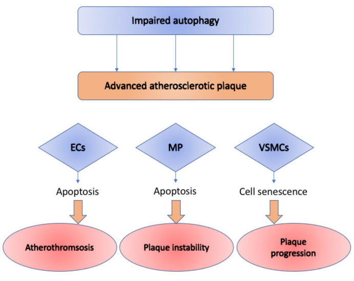
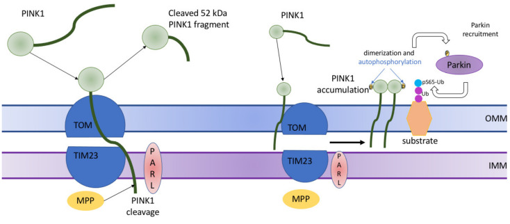
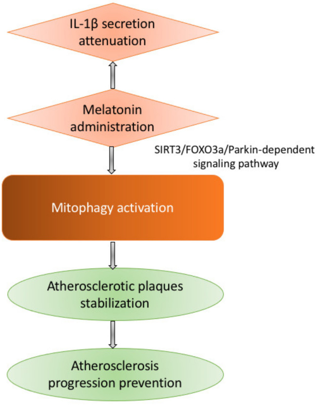
Cardiovascular disease (CVD) is one of the greatest health problems affecting people worldwide. Atherosclerosis, in turn, is one of the most common causes of cardiovascular disease. Due to the high mortality rate from cardiovascular diseases, prevention and treatment at the earliest stages become especially important. This requires developing a deep understanding of the mechanisms underlying the development of atherosclerosis. It is well-known that atherogenesis is a complex multi-component process that includes lipid metabolism disorders, inflammation, oxidative stress, autophagy disorders and mitochondrial dysfunction. Autophagy is a cellular control mechanism that is critical to maintaining health and survival. One of the specific forms of autophagy is mitophagy, which aims to control and remove defective mitochondria from the cell. Particularly defective mitophagy has been shown to be associated with atherogenesis. In this review, we consider the role of autophagy, focusing on a special type of it-mitophagy-in the context of its role in the development of atherosclerosis.

Keywords: atherosclerosis; autophagy; cardiovascular disease; mitochondria; mitochondrial dysfunction; mitophagy.


Conflict of interest statement

The authors declare no conflict of interest.

Figures

Figure 1
Figure 1
Impact of the impaired autophagy on cells of various types: ECs (endothelial cells), MP (macrophages), and VSMCs (vascular smooth muscle cells) in advanced atherosclerotic plaque. In Ecs, it causes cellular senescence and apoptosis, which leads to atherothrombosis; in MP—contributes to plaque instability through induced necrosis and apoptosis; in VSMCs—causes accelerated plaque formation through cellular senescence.
Figure 2
Figure 2
Brief description of PTEN-induced kinase 1 (PINK1)-mediated stimulation of mitophagy in normal and damaged mitochondria. In normal mitochondria (on the left), PINK1 is imported into mitochondria with the help of translocase of the outer membrane (TOM) complex and translocase of the inner membrane (TIM). At the IMM, PINK1 is initially processed by matrix processing peptidase (MPP), which removes PINK1’s N-terminal mitochondrial targeting signal, and then PINK1 is being proteolytically cleaved by the intermembrane serine protease presenilin-associated rhomboid-like protein (PARL), which cleaves the full-sized PINK1 form of 64 kDa into 60 kDa and 52 kDa fragments. In damaged mitochondria (on the right), PINK1 stores on the outer mitochondrial membrane of only injured mitochondria and is stabilized in a TOM complex (TOM7, TOM40, TOM70, TOM20, and TOM22). PINK1 mediates two various phosphorylations serving for the transformation of the autoinhibited E3-ubiquitin (Ub) ligase Parkin into an active phospho-Ub-dependent enzyme. Then, the direct phosphorylation of Parkin at S65 in the N-terminal Ubl domain occurs, increasing the activity of its E3 ligase. After that, PINK1 triggers the addition of phosphate onto S65 of Ub. This complex regulatory mechanism results in activation of the E3 ligase activity of Parkin, which allows it to ubiquitinate mitochondrial proteins through direct interaction with phospho-Ub conjugates on the mitochondria. OMM—outer mitochondrial membrane; IMM—inner mitochondrial membrane.
Figure 3
Figure 3
Melatonin administration affects atherosclerosis progression through an SIRT3/FOXO3a/Parkin-dependent signaling pathway. This leads to the stabilization of atherosclerotic plaques and, thus, prevents the further development of atherosclerosis. Also, melatonin reduces the IL-1β secretion.

References

    1. Galluzzi L., Vitale I., Aaronson S.A., Abrams J.M., Adam D., Agostinis P., Alnemri E.S., Altucci L., Amelio I., Andrews D.W., et al. Molecular mechanisms of cell death: Recommendations of the nomenclature committee on cell death 2018. Cell Death Differ. 2018;25:486–541. doi: 10.1038/s41418-017-0012-4. - DOI - PMC - PubMed
    1. Gatica D., Lahiri V., Klionsky D.J. Cargo recognition and degradation by selective autophagy. Nat. Cell. Biol. 2018;20:233–242. doi: 10.1038/s41556-018-0037-z. - DOI - PMC - PubMed
    1. Vural A., Kehrl J.H. Autophagy in macrophages: Impacting inflammation and bacterial infection. Scientifica. 2014;2014:825463. doi: 10.1155/2014/825463. - DOI - PMC - PubMed
    1. Tyrrell D.J., Goldstein D.R. Ageing and atherosclerosis: Vascular intrinsic and extrinsic factors and potential role of IL-6. Nat. Rev. Cardiol. 2021;18:58–68. doi: 10.1038/s41569-020-0431-7. - DOI - PMC - PubMed
    1. Lavandero S., Chiong M., Rothermel B.A., Hill J.A. Autophagy in cardiovascular biology. J. Clin. Investig. 2015;125:55–64. doi: 10.1172/JCI73943. - DOI - PMC - PubMed

Publication types

LinkOut - more resources