Review
. 2021 Feb 21;10(2):460.
doi: 10.3390/cells10020460.

The Pleiotropic Function of Human Sirtuins as Modulators of Metabolic Pathways and Viral Infections

Affiliations
Review

The Pleiotropic Function of Human Sirtuins as Modulators of Metabolic Pathways and Viral Infections

Mohammed Hamed Alqarni et al. Cells. .

Abstract

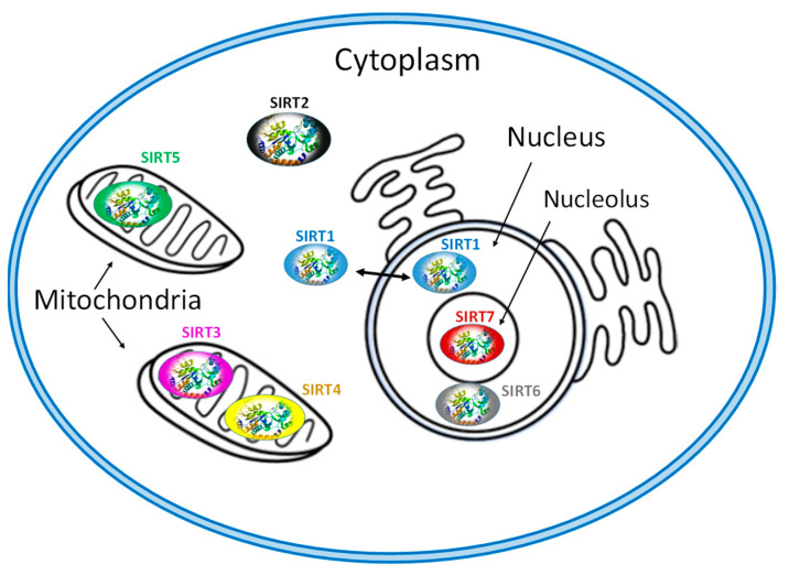
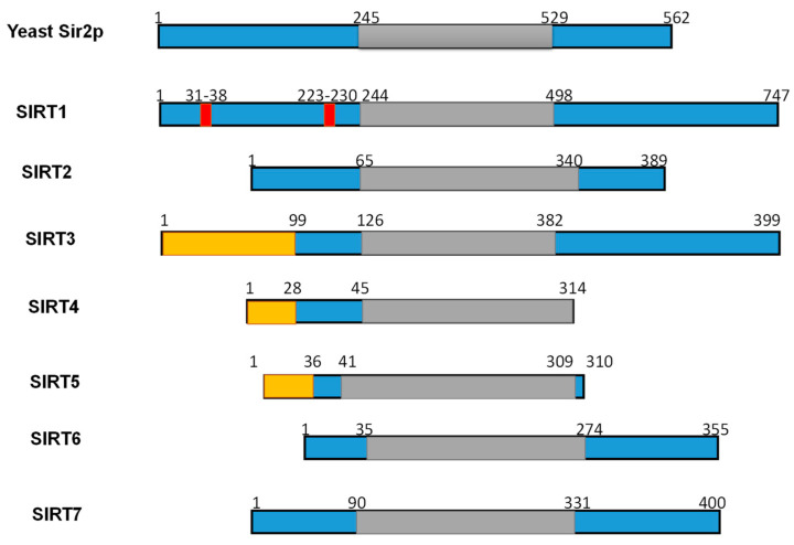
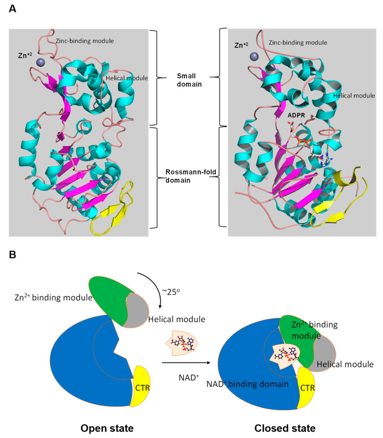
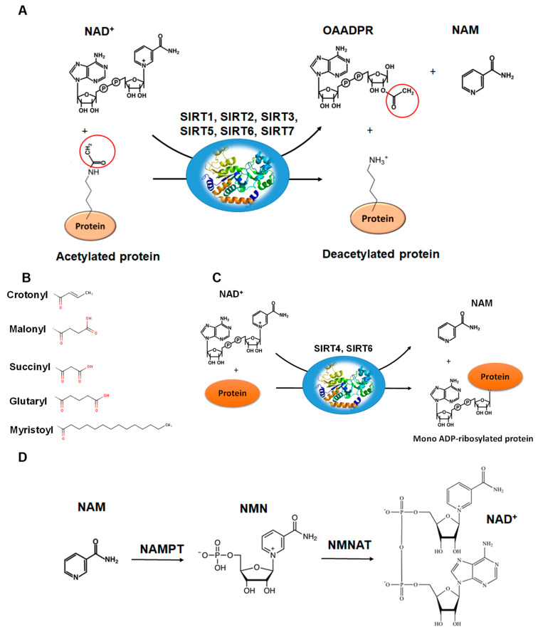
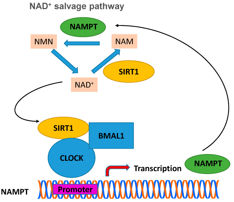
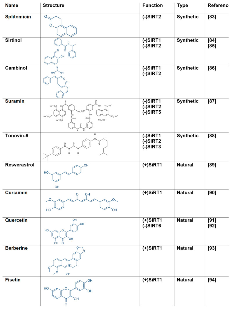
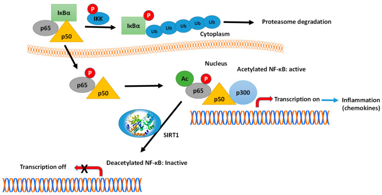
Sirtuins (SIRTs) are nicotinamide adenine dinucleotide-dependent histone deacetylases that incorporate complex functions in the mechanisms of cell physiology. Mammals have seven distinct members of the SIRT family (SIRT1-7), which play an important role in a well-maintained network of metabolic pathways that control and adapt the cell to the environment, energy availability and cellular stress. Until recently, very few studies investigated the role of SIRTs in modulating viral infection and progeny. Recent studies have demonstrated that SIRT1 and SIRT2 are promising antiviral targets because of their specific connection to numerous metabolic and regulatory processes affected during infection. In the present review, we summarize some of the recent progress in SIRTs biochemistry and their emerging function as antiviral targets. We also discuss the potential of natural polyphenol-based SIRT modulators to control their functional roles in several diseases including viral infections.

Keywords: Acetylation; COVID-19; NAD+; SIRT1; antiviral; infection; metabolism; sirtuins; virus.


Conflict of interest statement

The authors declare no conflict of interest.

Figures

Figure 1
Figure 1
Subcellular location of seven mammalian SIRTS (SIRT1-7).
Figure 2
Figure 2
Schematic overview of human SIRTs (SIRT1–7). Human SIRTs are aligned with yeast Sir2p. The conserved large catalytic domain is shown in grey. The nuclear localization sequences shown in red and the mitochondrial targeting sequences are shown in dark yellow. Numbers refer to amino acid residues in the proteins. Adapted from Ref. [9].
Figure 3
Figure 3
The tertiary structure of SIRT1. (A) The structure in open (left) conformation (SIRT1•CTR complex, PDB code 4IF6) and in closed (right) conformation (SIRT1•CTR•ADPR•Substrate complex, PDB code 4KXQ). The amino acid sequence of SIRT1 is organized into two independent domain. A cavity that is created between the two domains forms the active site. The C-terminal regulatory segment (CTR) is shown in yellow. The CTR binds at the lower edge of the larger NAD+-binding domain, complementing the central parallel β sheet of its Rossmann fold. The figure was created using the program PyMOL (www.pymol.org). (B) Cartoon model of the conformational changes of SIRT1 upon substrate and NAD+ analogue binding. The smaller domain undergoes a rotation with respect to the large domain. A cartoon representation of the apo SIRT1·CTR heterodimer (open state) and the SIRT1·CTR·NAD+·Substrate complex (closed state). Comparison of the open and closed structures revealed that the larger NAD+-binding domain does not undergo any major structural changes. The smaller domain rotates about 25°. The small domain (Zn2+-binding module and the helical module) rotates as a rigid body with only minor changes to the backbone and sidechain conformations. Adapted from Ref. [62].
Figure 4
Figure 4
The reactions catalyzed by SIRTs. (A) The general deacetylase reaction catalyzed by SIRT1,2,3,5,6,7. The natural substrates are NAD+, water and a protein containing acetylated lysine residue. The reaction products include nicotinamide (NAM), the deacetylated protein and the 2’-O-acetyl-ADPR (OAADPR) molecule. (B) Alternative acyl groups that can be deacylated by SIRT3,5,7. (C) The ADP-rebosyltransferase activity catalyzed by SIRT4 and SIRT6. (D) NAM can be used as precursor for the biosynthesis of NAD+ by the enzymes nicotinamide phosphoribosyltransferase (NAMPT) and nicotinamide mononucleotide adenylyltransferase (NMNAT).
Figure 5
Figure 5
The circadian clock machinery regulates the biosynthesis of NAD+ through control of the NAD+ salvage pathway. BMAL-1:CLOCK heterodimer binds to the promoter region of NAMPT to regulate the rhythmic transcription of this gene and thus the levels of NAD+. Expression of SIRT1 silences the expression of NAMPT (the rate-limiting enzyme in NAD+ biosynthesis), which leads to a decrease in the concentration of NAD+, diminishing the activity of SIRT1. When the activity of SIRT1 decreases significantly, then the activity of CLOCK:BMAL-1 begins to increase, which restores the expression levels of NAMPT and thus completes the cycle. NAMPT: nicotinamide phosphoribosyltransferase; CLOCK: basic helix-loop-helix-PAS transcription factor; BMAL1: Brain and muscle ARNT-like 1 protein; NMN: nicotinamide mononucleotide; NAM: nicotinamide.
Figure 6
Figure 6
The structure of natural and synthetic SIRT modulators [83,84,85,86,87,88,89,90,91,92,93,94].
Figure 7
Figure 7
Crystal structure of human SIRT1 in complex with resveratrol (shown in orange) and 7-amino-4-methylcoumarin containing peptide (shown in grey). The zinc ion is shown as a grey sphere. The figure was created using the program PyMOL (www.pymol.org).
Figure 8
Figure 8
Mechanism of NF-κB action and the role of SIRT1. In the inactivated state, the heterodimer NF-κB, composed by p65 (RelA) and p50 proteins, is located in the cytosol complexed with the inhibitory protein IκBα. A variety of extracellular signals can activate the enzyme IκB kinase (IKK), which phosphorylates the IκBα protein, leading to dissociation of the inhibitory protein IκBα from NF-κB. The phosphorylated IκBα is subjected to ubiquitination, leading to its degradation by the proteasome. The activated NF-κB is then translocated into the nucleus and interacts with specific sequences of DNA. The DNA/NF-κB complex then binds to coactivators (e.g., p300-CBP) and RNA polymerase, which transcribes downstream DNA into mRNA. SIRT1 suppresses NF-κB transcription factor by deacetylation of the p65 (RelA) subunit. Acetylation of NF-κB increases the transcription of proinflammatory mediators. Activators of SIRT1 can lead to repression of inflammation.

References

    1. Ren Z., He H., Zuo Z., Xu Z., Wei Z., Deng J. The role of different SIRT1-mediated signaling pathways in toxic injury. Cell Mol. Biol Lett. 2019;24:1–10. doi: 10.1186/s11658-019-0158-9. - DOI - PMC - PubMed
    1. Teixeira C.S.S., Cerqueira N.M.F.S.A., Gomes P., Sousa S.F. A Molecular Perspective on Sirtuin Activity. Int J. Mol. Sci. 2020;21:8609. doi: 10.3390/ijms21228609. - DOI - PMC - PubMed
    1. Wang Z., Guo W., Yi F., Zhou T., Li X., Feng Y., Guo Q., Xu H., Song X., Cao L. The Regulatory Effect of SIRT1 on Extracellular Microenvironment Remodeling. Int. J. Biol Sci. 2021;17:89–96. doi: 10.7150/ijbs.52619. - DOI - PMC - PubMed
    1. Soni S.K., Basu P., Singaravel M., Sharma R., Pandi-Perumal S.R., Cardinali D.P., Reiter R.J. Sirtuins and the circadian clock interplay in cardioprotection: Focus on sirtuin 1. Cell Mol. Life Sci. 2021 doi: 10.1007/s00018-020-03713-6. in press. - DOI - PMC - PubMed
    1. Ivy J.M., Klar A.J., Hicks J.B. Cloning and characterization of four SIR genes of Saccharomyces cerevisiae. Mol. Cell Biol. 1986;6:688–702. doi: 10.1128/MCB.6.2.688. - DOI - PMC - PubMed

Publication types

LinkOut - more resources