. 2021 Feb 11;11(2):457.
doi: 10.3390/nano11020457.

The Effect of a Polyester Nanofibrous Membrane with a Fibrin-Platelet Lysate Coating on Keratinocytes and Endothelial Cells in a Co-Culture System

Affiliations

The Effect of a Polyester Nanofibrous Membrane with a Fibrin-Platelet Lysate Coating on Keratinocytes and Endothelial Cells in a Co-Culture System

Andreu Blanquer et al. Nanomaterials (Basel). .

Abstract

Chronic wounds affect millions of patients worldwide, and it is estimated that this number will increase steadily in the future due to population ageing. The research of new therapeutic approaches to wound healing includes the development of nanofibrous meshes and the use of platelet lysate (PL) to stimulate skin regeneration. This study considers a combination of a degradable electrospun nanofibrous blend of poly(L-lactide-co-ε-caprolactone) and poly(ε-caprolactone) (PLCL/PCL) membranes (NF) and fibrin loaded with various concentrations of PL aimed at the development of bioactive skin wound healing dressings. The cytocompatibility of the NF membranes, as well as the effect of PL, was evaluated in both monocultures and co-cultures of human keratinocytes and human endothelial cells. We determined that the keratinocytes were able to adhere on all the membranes, and their increased proliferation and differentiation was observed on the membranes that contained fibrin with at least 50% of PL (Fbg + PL) after 14 days. With respect to the co-culture experiments, the membranes with fibrin with 20% of PL were observed to enhance the metabolic activity of endothelial cells and their migration, and the proliferation and differentiation of keratinocytes. The results suggest that the newly developed NF combined with fibrin and PL, described in the study, provides a promising dressing for chronic wound healing purposes.

Keywords: electrospun nanofibre; endothelial cells; fibrin; in vitro co-culture system; keratinocytes; platelet lysate; skin wound healing.


Conflict of interest statement

The authors declare no conflict of interest.

Figures

Figure 1
Figure 1
Schematic illustration of the single nanofibre coating procedure with fibrin and platelet lysate.
Figure 2
Figure 2
Schematic illustration of the co-culture system used in the experiments with the HaCaT cells and the human saphenous vein endothelial cells.
Figure 3
Figure 3
SEM images of the upper surface of a poly(L-lactide-co-ε-caprolactone)/poly(ε-caprolactone) (PLCL/PCL) nanofibrous membranes (A,B). Histogram of the PLCL/PCL fibre diameter distribution on the upper nanofibrous membranes’ (NF) surface (C). Confocal laser scanning microscope (CLSM) images measured in phosphate-buffered solution of the fluorescence-stained proteins on the upper surface of the NF modified from a solution containing Fbg and 50% of platelet lysate (PL) (NF50) (D,E). The proteins were stained with fluorescamin (green). The protein concentrations in the NF0, NF1, NF5, NF10, NF20, NF50, and NF100 prepared from solutions containing Fbg and 0%, 1%, 5%, 10%, 20%, 50% and 100% of PL (F). The superscript letters above the columns denote significant differences between the values that do not share the same superscript (p < 0.05).
Figure 4
Figure 4
Attenuated total reflection Fourier transform infrared spectroscopy (ATR-FTIR) analysis of the PLCL/PCL and the spectra of pure PCL and pure PLCL (A), and lyophilized PLCL/PCL nanofibrous membranes coated with fibrin (blend-Fb) and fibrin with 50% of platelet lysate (B).
Figure 5
Figure 5
HaCaT cells that adhered to the pure NF membranes (Ctrl, A) and to the NF membranes with fibrin assemblies containing 0–100 vol.% of PL, i.e., NF0 (B), NF1 (C), NF5 (D), NF10 (E), NF20 (F), NF50 (G) and NF100 (H) on day 1 following seeding. Images of the cells stained with phalloidin-TRITC (red); the cell nuclei were counterstained with Hoechst 33,258 (blue). Leica SPE microscope, obj. x 20. The cell population density (I) and the spreading area (J) of the HaCaT cells on the NF membranes on day 1 following seeding. The data are expressed as the mean ± standard error of the mean. The superscript letters above the columns denote significant differences between the NF membranes that do not share the same superscript (p < 0.05).
Figure 6
Figure 6
Metabolic activity/viability of the HaCaT cells that grew on the NF membranes with fibrin assemblies prepared using differing platelet lysate (PL) concentrations, i.e., 0, 1, 5, 10, 20, 50 and 100% PL (NF0, NF1, NF5, NF10, NF20, NF50, NF100, respectively). Pure NF membranes were used as the control sample (Ctrl). (A) HaCaT cells were grown on a single layer of NF for 1, 4, 7 and 14 days. The asterisks above the columns indicate significant differences compared to the control membrane (p < 0.05). (B) Comparison of the cells that grew on the NF membranes for 14 days (single) and the cells that grew on the NF membranes in a medium to which additional NF membranes with corresponding PL concentrations were added on day 7 following seeding (double). The asterisks above the columns indicate significant differences between the single and double layers (p < 0.05).
Figure 7
Figure 7
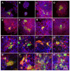
Immunofluorescence staining of the cytokeratin 10 (green) and cytokeratin 14 (red) in the HaCaT cells in the monoculture for the pure nanofibres (A,A’) and nanofibrous membranes with differing PL concentrations, i.e., NF0 (B,B’), NF1 (C,C’), NF5 (D,D’), NF10 (E,E’), NF20 (F,F’), NF50 (G,G’), NF100 (H,H’) (AH on day 7 and A’H’ on day 14). The cell nuclei were counterstained with Hoechst (blue). Leica SP8 confocal microscope, obj. × 20.
Figure 8
Figure 8
The metabolic activity of the HaCaT and HSVEC in the monocultures and co-cultures of these cell types. The metabolic activity of the HaCaT cells (A,B) that grew on the pure NF membranes (Ctrl), and on the NF membranes with fibrin assemblies prepared using various platelet lysate (PL) concentrations, i.e., 0%, 20% and 50% PL (NF0, NF20 and NF50, respectively). The metabolic activity of the human saphenous vein endothelial cells (HSVEC) (C,D) that grew on the well bottoms on a polystyrene well plate in the presence of the Ctrl, NF0, NF10, NF20 and NF50 membranes after 7 days (A,C) and 14 days (B,D) of cultivation. Each graph contains information on the cells that grew in the monoculture (white columns) and those co-cultured with the other cell type (grey columns). The superscript letters above the columns denote significant differences between the NF membranes that do not share the same superscript (p < 0.05).
Figure 9
Figure 9
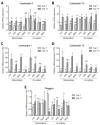
Quantification of the mRNA levels of the HaCaT in the monoculture and the co-culture with HSVEC. The relative expression of cytokeratin 5 (A), cytokeratin 14 (B), cytokeratin 1 (C), cytokeratin 10 (D) and filaggrin (E) on days 7 and 14 following seeding on the NF membranes. The target gene levels are expressed as relative values towards the reference B2M gene. The differing superscripts above the columns denote significant differences between the membranes that do not share the same superscript (p < 0.05) for each cell culture type separately (monoculture and co-culture). The lower case letters denote significant differences on day 7, the upper case letters on day 14.
Figure 10
Figure 10
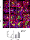
Differentiation of the HaCaT cells in the monoculture and co-culture on day 7 (AH) and on day 14 (A’H’). The HaCaT cells monocultured on the NF samples (AD,A’D’) and co-cultured with HSVECs (EH,E’H’) in the presence of the membranes as follows: Ctrl (A,E,A’,E’), NF0 (B,F,B’,F’), NF20 (C,G,C’,G’) and NF50 (D,H,D’,H’). The cells were stained with immunofluorescence for cytokeratin 10 (green) and for cytokeratin 14 (red). The cell nuclei were counterstained with Hoechst 33,258 (blue). Leica SP8 confocal microscope, obj x 20. Area (%) of the image covered with cells positive for cytokeratin 10 on the pure membranes (Ctrl), on the membranes with fibrin (NF0) and on the membranes with fibrin and 20% PL or 50% PL (NF20 and NF50) samples, analysed in the monoculture (white columns) and co-culture (grey columns) (I). The differing superscripts above the columns denote significant differences between the membranes that do not share the same superscript (p < 0.05).
Figure 11
Figure 11
The maturation of the HSVECs in the monoculture and co-culture on day 7. The HSVECs monocultured in polystyrene wells with NF membranes floating in the medium (AD) and co-cultured with HaCaT (EH) grown on the membranes. In both cases, the following membranes were used: Ctrl (A,E), NF0 (B,F), NF20 (C,G) and NF50 (D,H). The cells were stained with immunofluorescence for the von Willebrand factor (green); Olympus IX71 microscope (Olympus, Tokyo, Japan), DP80 digital camera (Olympus, Tokyo, Japan), obj. × 20. The number of HSVECs that transmigrated through the inserts towards the medium with the Ctrl, NF10, NF20, NF50 and NF100 samples over 4 h (I). The differing superscripts above the columns denote significant differences between the samples that do not share the same superscript (p < 0.05).

References

    1. Martinez-Zapata M., Martí-Carvajal A., Solà I., Expósito J., Bolíbar I., Rodríguez L., Garcia J., Zaror C. Autologous platelet-rich plasma for treating chronic wounds (Review) Cochrane Database Syst. Rev. 2016 doi: 10.1002/14651858.CD006899.pub3. - DOI - PMC - PubMed
    1. Sen C.K., Gordillo G.M., Roy S., Kirsner R., Lambert L., Hunt T.K., Gottrup F., Gurtner G.C., Longaker M.T. Human skin wounds: A major and snowballing threat to public health and the economy: PERSPECTIVE ARTICLE. Wound Repair Regen. 2009;17:763–771. doi: 10.1111/j.1524-475X.2009.00543.x. - DOI - PMC - PubMed
    1. Bacakova L., Zikmundova M., Pajorova J., Broz A., Filova E., Blanquer A., Matejka R., Stepanovska J., Mikes P., Jencova V., et al. Applications of Nanobiotechnology. IntechOpen; London, UK: 2020. Nanofibrous Scaffolds for Skin Tissue Engineering and Wound Healing Based on Synthetic Polymers.
    1. Jeong S.I., Jun I.D., Choi M.J., Nho Y.C., Lee Y.M., Shin H. Development of electroactive and elastic nanofibers that contain polyaniline and poly(L-lactide-co-ε-caprolactone) for the control of cell adhesion. Macromol. Biosci. 2008;8:627–637. doi: 10.1002/mabi.200800005. - DOI - PubMed
    1. Losi P., Barsotti M.C., Foffa I., Buscemi M., De Almeida C.V., Fabbri M., Gabbriellini S., Nocchi F., Ursino S., Urciuoli P., et al. In vitro human cord blood platelet lysate characterisation with potential application in wound healing. Int. Wound J. 2020;17:65–72. doi: 10.1111/iwj.13233. - DOI - PMC - PubMed

LinkOut - more resources