. 2021 Feb 11;11(2):462.
doi: 10.3390/nano11020462.

Quantum Dots as a Good Carriers of Unsymmetrical Bisacridines for Modulating Cellular Uptake and the Biological Response in Lung and Colon Cancer Cells

Affiliations

Quantum Dots as a Good Carriers of Unsymmetrical Bisacridines for Modulating Cellular Uptake and the Biological Response in Lung and Colon Cancer Cells

Joanna Pilch et al. Nanomaterials (Basel). .

Abstract

Nanotechnology-based drug delivery provides a promising area for improving the efficacy of cancer treatments. Therefore, we investigate the potential of using quantum dots (QDs) as drug carriers for antitumor unsymmetrical bisacridine derivatives (UAs) to cancer cells. We examine the influence of QD-UA hybrids on the cellular uptake, internalization (Confocal Laser Scanning Microscope), and the biological response (flow cytometry and light microscopy) in lung H460 and colon HCT116 cancer cells. We show the time-dependent cellular uptake of QD-UA hybrids, which were more efficiently retained inside the cells compared to UAs alone, especially in H460 cells, which could be due to multiple endocytosis pathways. In contrast, in HCT116 cells, the hybrids were taken up only by one endocytosis mechanism. Both UAs and their hybrids induced apoptosis in H460 and HCT116 cells (to a greater extent in H460). Cells which did not die underwent senescence more efficiently following QDs-UAs treatment, compared to UAs alone. Cellular senescence was not observed in HCT116 cells following treatment with both UAs and their hybrids. Importantly, QDgreen/red themselves did not provoke toxic responses in cancer or normal cells. In conclusion, QDs are good candidates for targeted UA delivery carriers to cancer cells while protecting normal cells from toxic drug activities.

Keywords: apoptosis; cellular senescence; cellular uptake; internalization; lung and colon cancer cells; quantum dots; unsymmetrical bisacridines.


Conflict of interest statement

The authors declare no conflict of interest.

Figures

Figure 1
Figure 1
Mean size, polydispersity index (PDI), and zeta potential of quantum dot (QD) nanocrystals, QD−UA (unsymmetrical bisacridine derivatives) hybrids dispersed in PBS buffer based on dynamic light scattering (DLS) studies (n = 5).
Figure 2
Figure 2
TEM images of QDs and their hybrids with UA compounds.
Figure 3
Figure 3
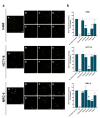
Cellular uptake of UAs, and QD−UA hybrids to cancer (H460 and HCT116) and normal (MRC-5 and CCD 841 CoN) cells for the time indicated and analyzed by Confocal Laser Scanning Microscope (CLSM). Mean Fluorescence Intensity (MFI) values of UAs and QDs−UAs were determined using ImageJ software. Data are expressed as the mean ± standard deviation of three independent experiments. * p < 0.05, ** p < 0.01, *** p < 0.001—statistically significant differences between the MFI of UAs in the cells incubated with UAs alone and QD−UA hybrids (Student’s t-test).
Figure 4
Figure 4
(a) The influence of different endocytosis inhibitors on the internalization of QDgreen-C-2028 in cancer and normal cells. H460, HCT116, and MRC-5 cells were preincubated with (A) drug-free medium (no inhibitor), (B) at 4 °C, (C) 5 µM Cytochalasin D, (D) 30 µM Amiloride, (E) 80 µM Dynasore, (F) 25 µM Pitstop 2, and (G) 1.5 µM Filipin III for 30 min, followed by further incubation with QDgreen-C-2028 for 4 h. The internalization of QDgreen-C-2028 in cells was explored by CLSM. Scale bar 10 µm. (b) MFI values of the panel (a) were determined using ImageJ software and normalized to control (cells treated QDgreen-C-2028 without inhibitor). Data are expressed as the mean ± standard deviation of four independent experiments. * p < 0.05, ** p < 0.01, *** p < 0.001—statistically significant differences between the MFI of UAs and QDs in the cells incubated with inhibitors and control cells (no inhibitor) (Student’s t-test).
Figure 5
Figure 5
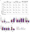
Cell cycle analysis of H460, HCT116, MRC-5, and CCD 841 CoN cells following treatment with QDs, UAs, and QD−UA hybrids for the time indicated. Cells were fixed in ethanol and stained with propidium iodide (PI), and their DNA content was measured by flow cytometry. (a) Representative plots of cells treated with selected QDs, UAs, and QD−UA hybrids. (b) Quantification of data shows the cell distributions in the sub-G1, G1, and G2/M phases of the cell cycle. Data represented the averages of three independent experiments. * p < 0.05, ** p < 0.01, *** p < 0.001 indicates statistically significant differences between fraction of cells incubated with UAs alone and QD−UA hybrids (Student’s t-test).
Figure 6
Figure 6
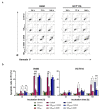
Flow cytometry analysis of phosphatidylserine externalization by Annexin V/propidium iodide (PI) assay in H460 and HCT116 cells. (a) Representative plots of cells treated with selected QDs, UAs, and QD−UA hybrids. (b) Quantification of data expressed as the percentage of late apoptotic cells (A+/PI+). Data represented the averages of three independent experiments. * p < 0.05, ** p < 0.01, *** p < 0.001 indicate statistically significant differences between the fraction of late apoptotic cells incubated with UAs alone and QD−UA hybrids (Student’s t-test). Bottom left quadrant represents live cells (Annexin V negative, PI negative); bottom right quadrant—early apoptotic cells (Annexin V positive, PI negative); top right quadrant—late apoptotic cells (Annexin V positive, PI positive); top left quadrant—primary necrotic cells (Annexin V negative, PI positive).
Figure 7
Figure 7
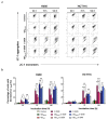
Analysis of the changes in mitochondrial membrane potential (ΔΨm) in H460 and HCT116 cells following treatment with QDs, UAs, and QD−UA hybrids for 24, 72, and 144 h. ΔΨm was measured by flow cytometry using JC-1 dye. (a) Representative plots of cells treated with selected QDs, UAs, and QD−UA hybrids. (b) Quantification of data expressed as the percentage of cells with decreased ΔΨm. Data represented the averages of three independent experiments. * p < 0.05, ** p < 0.01, *** p < 0.001 indicate statistically significant differences between the percentage of cells with decreased ΔΨm incubated with UAs alone and QD−UA hybrids (Student’s t-test).
Figure 8
Figure 8
Cellular senescence of H460 and HCT116 cancer cells following treatment with QDs, UAs, and QD−UA hybrids for the time indicated. Senescence-associated β-galactosidase activities were assessed by X-gal staining using a light microscope. (a) Representative images of cells treated with selected UAs and QD−UA hybrids. (b) Quantification of data expressed as the percentage of SA-β-gal positive cells in H460 cells. (c) Representative images of cells treated with QDs in H460 and HCT116 cells. Data represented the averages of three independent experiments. * p < 0.05, ** p < 0.01, *** p < 0.001 indicate statistically significant differences between the percentage of SA-β-gal positive cells incubated with UAs alone and QD−UA hybrids (Student’s t-test). The scale bar 50 µm.

References

    1. Zhang Y., Huang F., Ren C., Yang L., Liu J., Cheng Z., Chu L., Liu J. Targeted Chemo-Photodynamic Combination Platform Based on the DOX Prodrug Nanoparticles for Enhanced Cancer Therapy. ACS Appl. Mater. Interfaces. 2017;9:13016–13028. doi: 10.1021/acsami.7b00927. - DOI - PubMed
    1. Chakraborty S., Rahman T. The difficulties in cancer treatment. Ecancermedicalscience. 2012;6 doi: 10.3332/ecancer.2012.ed16. - DOI - PMC - PubMed
    1. Liang B., Li N., Zhang S., Qi A., Feng J., Jing W., Shi C., Ma Z., Gao S. Idarubicin-loaded methoxy poly(ethylene glycol)-b-poly(L-lactide-co-glycolide) nanoparticles for enhancing cellular uptake and promoting antileukemia activity. Int. J. Nanomed. 2019;14:543–556. doi: 10.2147/IJN.S190027. - DOI - PMC - PubMed
    1. Kübler E., Albrecht H. Large set data mining reveals overexpressed GPCRs in prostate and breast cancer: Potential for active targeting with engineered anti-cancer nanomedicines. Oncotarget. 2018;9:24882–24897. doi: 10.18632/oncotarget.25427. - DOI - PMC - PubMed
    1. Senapati S., Mahanta A.K., Kumar S., Maiti P. Controlled drug delivery vehicles for cancer treatment and their performance. Signal Transduct. Target. Ther. 2018;3:7. doi: 10.1038/s41392-017-0004-3. - DOI - PMC - PubMed

LinkOut - more resources