. 2021 Feb 11;13(2):283.
doi: 10.3390/v13020283.

Elephant Endotheliotropic Herpesvirus Is Omnipresent in Elephants in European Zoos and an Asian Elephant Range Country

Affiliations

Elephant Endotheliotropic Herpesvirus Is Omnipresent in Elephants in European Zoos and an Asian Elephant Range Country

Tabitha E Hoornweg et al. Viruses. .

Abstract

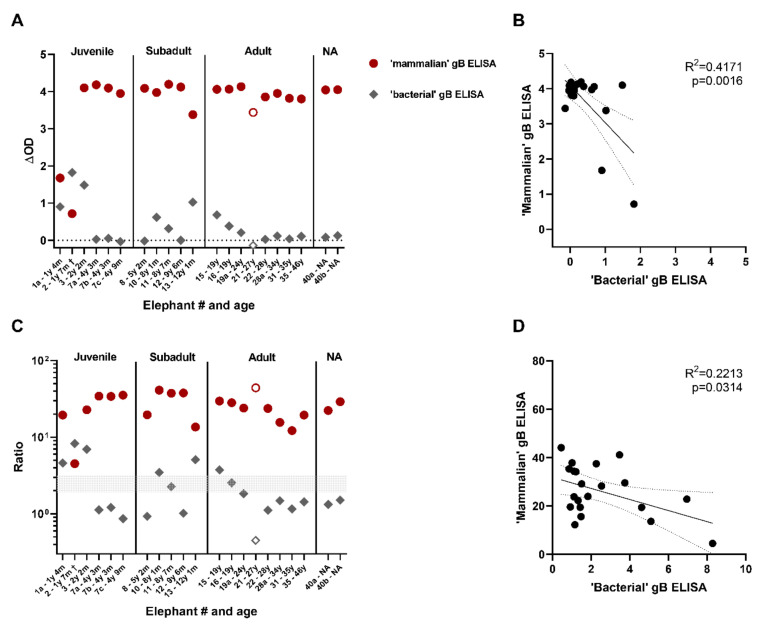
Elephant endotheliotropic herpesviruses (EEHVs) may cause acute, often lethal, hemorrhagic disease (EEHV-HD) in young elephants. Prevalence of EEHV in different elephant populations is still largely unknown. In order to improve diagnostic tools for the detection of EEHV infections and to obtain insight into its spread among elephants, we developed novel ELISAs based on EEHV1A gB and gH/gL. Performance of the ELISAs was assessed using sera from 41 European zoo elephants and 69 semi-captive elephants from Laos, one of the Asian elephant range countries. Sera from all (sub)adult animals tested (≥5 years of age) showed high reactivity with both gB and gH/gL, indicating that EEHV prevalence has been highly underestimated so far. Reactivity towards the antigens was generally lower for sera of juvenile animals (1 > 5 years). Only one (juvenile) animal, which was sampled directly after succumbing to EEHV-HD, was found to be seronegative for EEHV. The two other EEHV-HD cases tested showed low antibody levels, suggesting that all three cases died upon a primary EEHV infection. In conclusion, our study suggests that essentially all (semi-)captive (sub)adult elephants in European zoos and in Laos carry EEHV, and that young elephants with low antibody levels are at risk of dying from EEHV-HD.

Keywords: EEHV; elephant; gB; gH/gL; herpesviruses; immunoserology.


Conflict of interest statement

The authors declare no conflict of interest. Funders had no role in the study design, data collection and analysis, decision to publish, or preparation of the manuscript.

Figures

Figure 1
Figure 1
Production of recombinant EEHV1A gB, gH/gL and gL using the HEK293T expression system. (A) Schematic representation of the recombinant (i) gB-ST, (ii) gH-ST, (iii) gL-ST and (iv) gL-HT proteins. Amino acid mutations introduced into the two putative fusion loops (light green) and furin cleavage site (dark green) of gB are indicated as black bars. (B) Representative Western Blots showing the cell lysate (c) and secreted (s) fractions of recombinant EEHV1A gB, gH, gL, gH/gL and the IAV HA control produced in HEK293T cells. (C) Western Blot of the cell lysate (c) and secreted (s) fractions harvested from a gH-ST/gL-ST co-transfection subjected to SDS-PAGE under reducing and non-reducing conditions. Proteins were deglycosylated by PNGaseF prior to electrophoresis. (D) Gelcode Blue-stained purified EEHV1A gB- ST, gH-ST/gL-HT and gL-ST. Samples were deglycosylated by PNGaseF prior to electrophoresis to facilitate analysis. Molecular mass markers are indicated on the left side of the gels. EEHV = Elephant endotheliotropic herpesvirus
Figure 2
Figure 2
Antigen and serum dilution curves for the novel gB, gH/gL and gL ELISAs. Dilution ranges (40–1.25 ng/well) of (A) gB, (B) gH/gL, and (C) gL. (DF) Serum dilution ranges assayed using optimal antigen dilution determined in (AC), namely (D) 5 ng gB, (E) 40 ng gH/gL and (F) 40 ng gL. Dilution ranges were performed either single or in duplo, and results were depicted as the ΔOD (signal in antigen-coated well—signal in uncoated well). Elephant sera used are indicated by elephant number, species (Asian elephant = Elephas maximus (EM); African elephant = Loxodonta africana (LA)) and age. Samples indicated by a † were taken perimortem from lethal EEHV-HD cases.
Figure 3
Figure 3
Assessment of EEHV seropositivity in elephants living in European zoos. ΔOD values of 50 elephant sera from 41 individual elephants obtained in the gB (A), gH/gL ((B)—blue squares) and gL ((B)—green triangles) ELISAs. Sera are identified by elephant number and age and ordered based on age at time of sampling on the x-axis. Longitudinal or paired samples of individual elephants are identified by the individual elephant number followed by a character (either a, b, or c). Samples from Asian elephants (Elephas maximus = EM) are shown by closed symbols and those from African elephants (Loxodonta Africana = LA) by open symbols. Samples indicated by a † were taken perimortem from lethal EEHV-HD cases. The majority of samples were tested once, while 8 (gB), 10 (gH/gL), and 4 (gL) sera were assayed multiple times (range 2– 10 times) with comparable results. For these samples, standard deviations are indicated, which are only visible when they exceed the size of the symbol used. (C) ΔOD values of the longitudinal or paired serum samples of eight individual elephants. (D) ΔOD values of individual (sub)adult elephants divided by elephant species. For elephants with longitudinal samples available, the serum of the last sampling date was selected for analysis. Significance was tested by Mann–Whitney-test using Graphpad Prism: *** = p ≤ 0.001. OD = optical density
Figure 4
Figure 4
Comparison of the novel ‘mammalian’ gB ELISA to the ‘bacterial’ gB ELISA. (A) ΔOD values of 21 sera (of 18 individual elephants) tested using both the novel ‘mammalian’ gB ELISA (red circles) and a previously published ‘bacterial’ gB ELISA (grey diamonds) [12]. Closed symbols indicate Asian elephants (EM, n = 17) while open symbols African elephants (LA, n = 1). Samples indicated by a † were taken perimortem from lethal EEHV-HD cases. Sera were tested at both 1:100 and 1:200 dilutions in the ‘bacterial’ gB ELISA; the most optimal (highest) ΔOD ratio is shown. (B) Correlation between the ΔOD values of the ‘mammalian’ and ‘bacterial’ gB ELISA shown in (A). (C) Antigen-specific OD/background OD-ratio calculated for ELISAs. Sera were tested at both 1:100 and 1:200 dilutions in the bacterial gB ELISA; the most optimal (highest) ΔOD ratio is shown. Symbols as described in (A). (D) Correlation between the antigen-specific OD/background OD-ratio’s shown in (C). Correlations were analyzed using GraphPad Prism.
Figure 5
Figure 5
Reactivity of five EEHV1A gB peptide-specific rabbit antisera towards mammalian and bacterial produced gB. (A) Positions of the five gB peptides to which rabbit antisera were generated are indicated [12]. (B) ΔOD values for the gB peptide-specific rabbit sera measured in the mammalian gB (red circles) and bacterial gB (grey diamonds) ELISAs. Sera were tested at least once in duplicate at a 1:100 dilution. Representative results are shown.
Figure 6
Figure 6
Results obtained in the gB and gH/gL ELISAs for 69 serum samples from individual Asian elephants living under human care in Laos. Samples are plotted based on age at sampling. SA = subadult, NA = information on age not available.

References

    1. Howard L., Schaftenaar W. Fowler’s Zoo and Wild Animal Medicine Current Therapy. Vol. 9 Elsevier; Saunders, PA, USA: 2018. Elephant Endotheliotropic Herpesvirus.
    1. Jeffrey A., Evans T.S., Molter C., Howard L.L., Ling P., Goldstein T., Gilardi K. Noninvasive sampling for detection of elephant endotheliotropic herpesvirus and genomic DNA in Asian (Elephas maximus) and African (Loxodonta africana) elephants. J. Zoo Wildl. Med. 2020;51:433–437. doi: 10.1638/2019-0112. - DOI - PMC - PubMed
    1. Long S.Y., Latimer E.M., Hayward G.S. Review of Elephant Endotheliotropic Herpesviruses and Acute Hemorrhagic Disease. ILAR J. 2016;56:283–296. doi: 10.1093/ilar/ilv041. - DOI - PMC - PubMed
    1. Boonprasert K., Punyapornwithaya V., Tankaew P., Angkawanish T., Sriphiboon S., Titharam C., Brown J.L., Somgird C. Survival analysis of confirmed elephant endotheliotropic herpes virus cases in Thailand from 2006–2018. PLoS ONE. 2019;14:e0219288. doi: 10.1371/journal.pone.0219288. - DOI - PMC - PubMed
    1. Mahato G., Sarma K.K., Pathak D.C., Barman N.N., Gogoi P., Dutta M., Basumatary P. Endotheliotropic herpesvirus infection in Asian elephants (Elephas maximus) of Assam, India. Vet. World. 2019;12:1790–1796. doi: 10.14202/vetworld.2019.1790-1796. - DOI - PMC - PubMed

Publication types

MeSH terms

LinkOut - more resources