

Synergistic Roles of Curcumin in Sensitising the Cisplatin Effect on a Cancer Stem Cell-Like Population Derived from Non-Small Cell Lung Cancer Cell Lines
- PMID: 33670440
- PMCID: PMC7922800
- DOI: 10.3390/molecules26041056
Synergistic Roles of Curcumin in Sensitising the Cisplatin Effect on a Cancer Stem Cell-Like Population Derived from Non-Small Cell Lung Cancer Cell Lines
Abstract
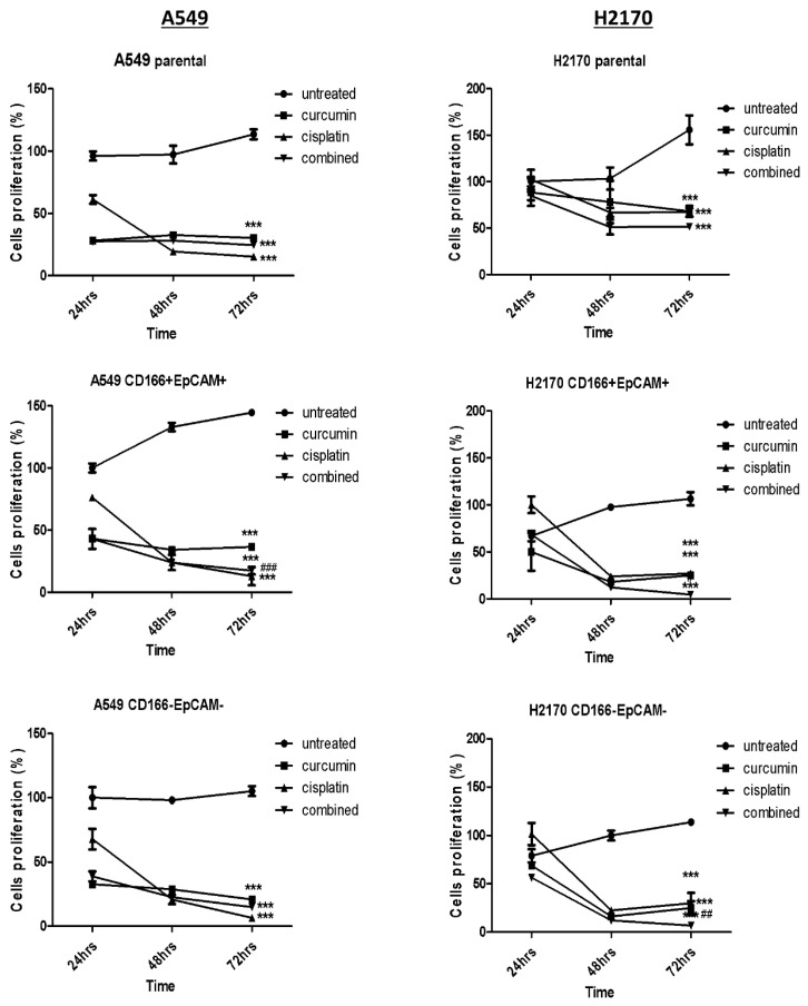
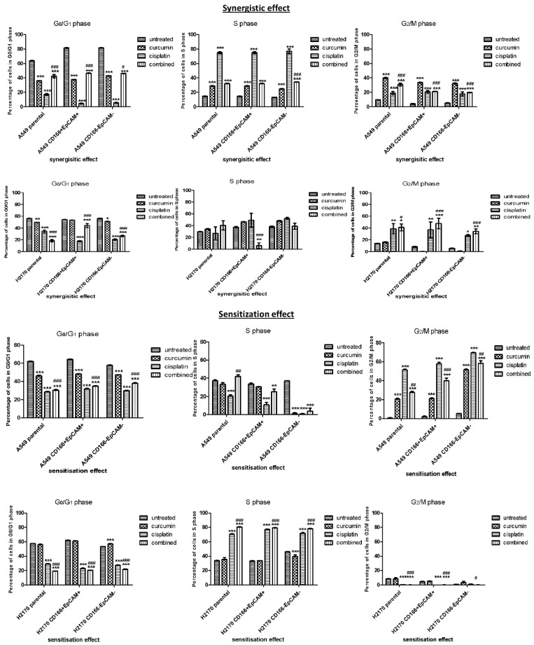
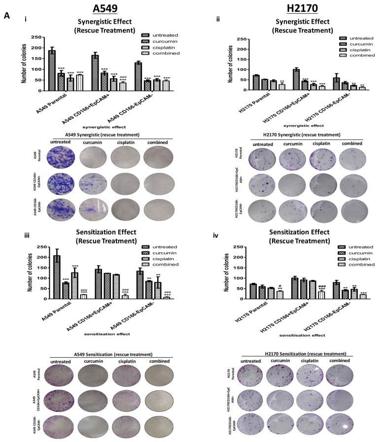
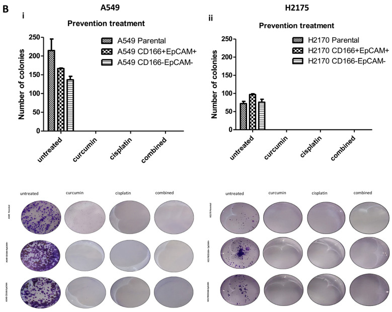
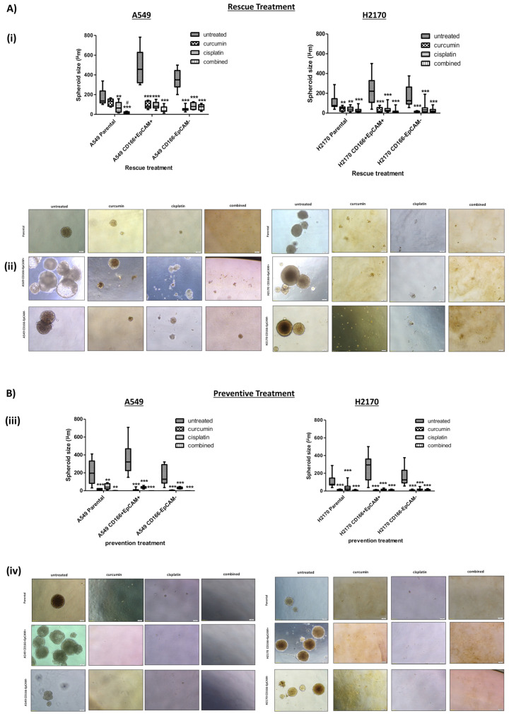
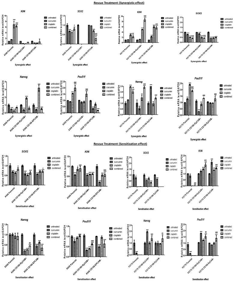
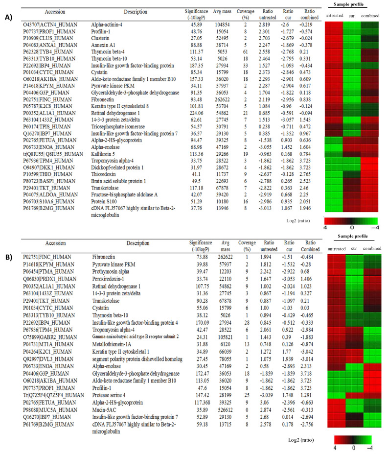
Cancer stem cells (CSCs) represent a small subpopulation within a tumour. These cells possess stem cell-like properties but also initiate resistance to cytotoxic agents, which contributes to cancer relapse. Natural compounds such as curcumin that contain high amounts of polyphenols can have a chemosensitivity effect that sensitises CSCs to cytotoxic agents such as cisplatin. This study was designed to investigate the efficacy of curcumin as a chemo-sensitiser in CSCs subpopulation of non-small cell lung cancer (NSCLC) using the lung cancer adenocarcinoma human alveolar basal epithelial cells A549 and H2170. The ability of curcumin to sensitise lung CSCs to cisplatin was determined by evaluating stemness characteristics, including proliferation activity, colony formation, and spheroid formation of cells treated with curcumin alone, cisplatin alone, or the combination of both at 24, 48, and 72 h. The mRNA level of genes involved in stemness was analysed using quantitative real-time polymerase chain reaction. Liquid chromatography-mass spectrometry was used to evaluate the effect of curcumin on the CSC niche. A combined treatment of A549 subpopulations with curcumin reduced cellular proliferation activity at all time points. Curcumin significantly (p < 0.001) suppressed colonies formation by 50% and shrank the spheroids in CSC subpopulations, indicating inhibition of their self-renewal capability. This effect also was manifested by the down-regulation of SOX2, NANOG, and KLF4. Curcumin also regulated the niche of CSCs by inhibiting chemoresistance proteins, aldehyde dehydrogenase, metastasis, angiogenesis, and proliferation of cancer-related proteins. These results show the potential of using curcumin as a therapeutic approach for targeting CSC subpopulations in non-small cell lung cancer.
Keywords: cisplatin; curcumin; lung cancer stem cells; non-small cell lung cancer; preventive; sensitisation.
Conflict of interest statement
The authors declare that the research was conducted in the absence of any commercial or financial relationships that could be construed as a potential conflict of interest.
Figures
Similar articles
-
Curcumin improves the efficacy of cisplatin by targeting cancer stem-like cells through p21 and cyclin D1-mediated tumour cell inhibition in non-small cell lung cancer cell lines.Oncol Rep. 2016 Jan;35(1):13-25. doi: 10.3892/or.2015.4371. Epub 2015 Nov 2. Oncol Rep. 2016. PMID: 26531053 Free PMC article.
-
Novel triple‑positive markers identified in human non‑small cell lung cancer cell line with chemotherapy-resistant and putative cancer stem cell characteristics.Oncol Rep. 2018 Aug;40(2):669-681. doi: 10.3892/or.2018.6461. Epub 2018 May 24. Oncol Rep. 2018. PMID: 29845263 Free PMC article.
-
Jorunnamycin A Suppresses Stem-Like Phenotypes and Sensitizes Cisplatin-Induced Apoptosis in Cancer Stem-Like Cell-Enriched Spheroids of Human Lung Cancer Cells.Mar Drugs. 2021 May 3;19(5):261. doi: 10.3390/md19050261. Mar Drugs. 2021. PMID: 34063628 Free PMC article.
-
The Role of Cancer Stem Cells in Recurrent and Drug-Resistant Lung Cancer.Adv Exp Med Biol. 2016;890:57-74. doi: 10.1007/978-3-319-24932-2_4. Adv Exp Med Biol. 2016. PMID: 26703799 Review.
-
Extracellular vesicles in non-small cell lung cancer stemness and clinical applications.Front Immunol. 2024 May 3;15:1369356. doi: 10.3389/fimmu.2024.1369356. eCollection 2024. Front Immunol. 2024. PMID: 38765006 Free PMC article. Review.
Cited by
-
Combination Chemotherapy with Selected Polyphenols in Preclinical and Clinical Studies-An Update Overview.Molecules. 2023 Apr 26;28(9):3746. doi: 10.3390/molecules28093746. Molecules. 2023. PMID: 37175156 Free PMC article.
-
Redefining Chemoresistance: Natural Bioactives as Molecular Modulators at the Cancer-Tumor Microenvironment Interface.Int J Mol Sci. 2025 Aug 20;26(16):8037. doi: 10.3390/ijms26168037. Int J Mol Sci. 2025. PMID: 40869364 Free PMC article. Review.
-
Curcumin and Its Analogs in Non-Small Cell Lung Cancer Treatment: Challenges and Expectations.Biomolecules. 2022 Nov 4;12(11):1636. doi: 10.3390/biom12111636. Biomolecules. 2022. PMID: 36358986 Free PMC article. Review.
-
Antitumor Therapy Targeting the Tumor Microenvironment.J Oncol. 2023 Mar 3;2023:6886135. doi: 10.1155/2023/6886135. eCollection 2023. J Oncol. 2023. PMID: 36908706 Free PMC article. Review.
-
Anti-Cancer Mechanisms of Diarylpentanoid MS17 (1,5-Bis(2-hydroxyphenyl)-1,4-pentadiene-3-one) in Human Colon Cancer Cells: A Proteomics Approach.Int J Mol Sci. 2024 Mar 20;25(6):3503. doi: 10.3390/ijms25063503. Int J Mol Sci. 2024. PMID: 38542474 Free PMC article.
References
MeSH terms
Substances
Grants and funding
LinkOut - more resources
Full Text Sources
Other Literature Sources
Medical
Research Materials
