. 2021 Feb 18;11(2):302.
doi: 10.3390/biom11020302.

New Insights into the Antimicrobial Action of Cinnamaldehyde towards Escherichia coli and Its Effects on Intestinal Colonization of Mice

Affiliations

New Insights into the Antimicrobial Action of Cinnamaldehyde towards Escherichia coli and Its Effects on Intestinal Colonization of Mice

Wellison A Pereira et al. Biomolecules. .

Abstract

Escherichia coli is responsible for cases of diarrhea around the world, and some studies have shown the benefits of cinnamaldehyde in the treatment of bacterial disease. Therefore, the objective of this study was to evaluate the effects of cinnamaldehyde in mice colonized by pathogenic E. coli, as well as to provide more insights into its antimicrobial action mechanism. After determination of minimum inhibitory (MIC) and minimum bactericidal (MBC) concentrations, the interference of cinnamaldehyde in macromolecular pathways (synthesis of DNA, RNA, protein, and cell wall) was measured by incorporation of radioisotopes. The anti-adhesive properties of cinnamaldehyde towards E. coli 042 were evaluated using human epithelial type 2 (HEp-2) cells. Intestinal colonization was tested on mice, and the effect of cinnamaldehyde on Tenebrio molitor larvae. Cinnamaldehyde showed MIC and MBC values of 780 μg/mL and 1560 μg/mL, respectively; reduced the adhesion of E. coli 042 on HEp-2 cells; and affected all the synthetic pathways evaluated, suggesting that compost impairs the membrane/cell wall structure leading bacteria to total collapse. No effect on the expression of genes related to the SOS pathway (sulA and dinB1) was observed. The compound did not interfere with cell viability and was not toxic against T. molitor larvae. In addition, cinnamaldehyde-treated mice exhibited lower levels of colonization by E. coli 042 than the untreated group. Therefore, the results show that cinnamaldehyde is effective in treating the pathogenic E. coli strain 042 and confirm it as a promising lead molecule for the development of antimicrobial agents.

Keywords: cinnamaldehyde; intestinal colonization; natural products.


Conflict of interest statement

All authors: No reported conflicts.

Figures
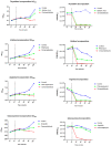
Figure 1
Figure 1
Effects of cinnamaldehyde on the assembly of the main bacterial structures by incorporating the radioactive precursors thymidine, uridine, arginine, and glucosamine that are essential for the synthesis of DNA, RNA, proteins, and cell wall, respectively. The cultures were treated with cinnamaldehyde or antibiotic after 35 min. The samples were collected at determined periods (15, 35, 50, 60, 80, 100, and 120 min) and transferred to a tube containing 0.375 μCi of each precursor. The sample was allowed to stand, according to the specific time of incorporation of each precursor. Thus, the value obtained by reading the radioactivity was subtracted from the value generated by the O.D., and determined the final result of the incorporation.
Figure 2
Figure 2
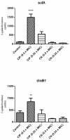
Expression of sulA and dinB1, genes linked to the SOS response. The test was performed using E. coli sulA::lacZ (ALO 4696) and E. coli dinB1::lacZ (ALO 562). The strains were grown in LB broth until OD600 of 0.1 was reached. Strains were incubated with ciprofloxacin as positive control and cinnamaldehyde. Then the supernatant was added to 1 mL of Zeta buffer containing ONPG (substrate for the enzyme β-Galactosidase). The tubes were again incubated in a 30 °C water bath with shaking and the time taken for each sample until the color change was verified. **** p < 0.0001 when compared to the other groups. ** p < 0.001 when compared to the other groups.
Figure 3
Figure 3
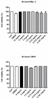
Cell viability assay (MTT) with Vero and HEp-2 cells incubated with cinnamaldehyde at different concentrations for 48 h. To evaluate a possible cytotoxic effect of cinnamaldehyde, the compound at various concentrations was incubated with VERO or Hep-2 cells for 48 h. Then 100 µL MTT (3- (4,5-dimethylthiazol-2-yl) -2,5-diphenyltetrazolium bromide) was added to each well and the plates were incubated for 3 h (37 °C and 5% CO2); after, 2 µL of dimethyl sulfoxide (DMSO) was added. The contents of each well were subjected to absorbance determination at a wavelength of 550 nm.
Figure 4
Figure 4
Survival curve with Tenebrio molitor larvae treated with PBS or cinnamaldehyde. Larvae (~100 mg) were randomly assigned to experimental groups (n = 10/group), and then treated with injection of 10 μL cinnamaldehyde or PBS. After injections, the larvae were incubated at 37 °C and the mortality rate was observed for seven days after infection.
Figure 5
Figure 5
E. coli 042 adhesion test on HEp-2 cells exposed to cinnamaldehyde (600 µg/mL). The cells were stained with May–Grünwald / Giemsa solution and visualized by 1,000-fold light microscopy. (A) HEp-2 cell control, (B) EAEC 042 strain aggregate adhesion pattern, (C) strain 042 adhesion pattern after cinnamaldehyde treatment (600 µg/mL).
Figure 6
Figure 6
Colony forming units isolated from feces from animals colonized by EAEC 042 and treated with cinnamaldehyde. Suspensions of E. coli strain 042 were prepared to a final concentration of 5 × 10³ CFU/mL. The inoculum (200 μL) was administered orally, and fresh feces were collected daily up to 15 days after infection. Feces were weighed, homogenized in 1x sterile PBS (pH 7.4), and serial dilutions were seeded in MacConkey Agar containing streptomycin (100 μg/mL) for CFU counting.

Similar articles

Cited by

References

    1. Taghadosi R., Shakibaie M.R., Hosseini-Nave H. Antibiotic resistance, ESBL genes, integrons, phylogenetic groups and MLVA profiles of Escherichia coli pathotypes isolated from patients with diarrhea and farm animals in south-east of Iran. Comp. Immunol. Microbiol. Infect. Dis. 2019;63:117–126. doi: 10.1016/j.cimid.2019.01.004. - DOI - PubMed
    1. Tharwat N., El-Sherif R., Elnagdy S., Marzaban R., Amer S. Virulent Escherichia coli strains among Egyptian patients with acute diarrhoea versus urinary tract infection, and their antibiotic susceptibility. Arab J. Gastroenterol. 2019;20:74–80. doi: 10.1016/j.ajg.2019.01.003. - DOI - PubMed
    1. Verma S., Venkatesh V., Kumar R., Kashyap S., Kumar M., Maurya A.K., Singh M. Etiological agents of diarrhea in hospitalized pediatric patients with special emphasis on diarrheagenic Escherichia coli in North India. J. Lab. Phys. 2019;11:68. doi: 10.4103/JLP.JLP_123_18. - DOI - PMC - PubMed
    1. Rasko D.A., Webster D.R., Sahl J.W., Bashir A., Boisen N., Scheutz F., Paxinos E.E., Sebra R., Chin C.S. Origins of the E. coli strain causing an outbreak of hemolytic-uremic syndrome in Germany. Nejm. 2011;365:709–717. doi: 10.1056/NEJMoa1106920. - DOI - PMC - PubMed
    1. Jensen B.H., Olsen K.E., Struve C., Krogfelt K.A., Petersen A.M. Epidemiology and clinical manifestations of enteroaggregative Escherichia coli. Clin. Microbiol. Rev. 2014;27:614–630. doi: 10.1128/CMR.00112-13. - DOI - PMC - PubMed

Publication types

LinkOut - more resources