. 2021 Feb 18;22(4):2010.
doi: 10.3390/ijms22042010.

BMP Receptor Inhibition Enhances Tissue Repair in Endoglin Heterozygous Mice

Affiliations

BMP Receptor Inhibition Enhances Tissue Repair in Endoglin Heterozygous Mice

Wineke Bakker et al. Int J Mol Sci. .

Abstract

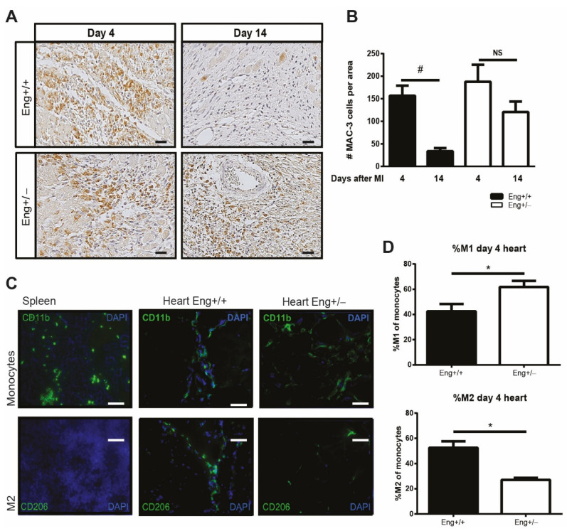
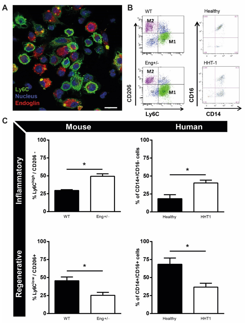
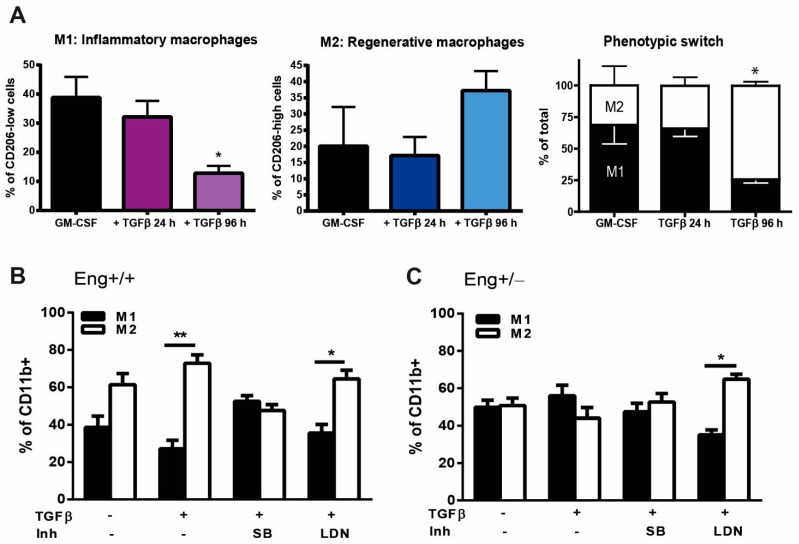
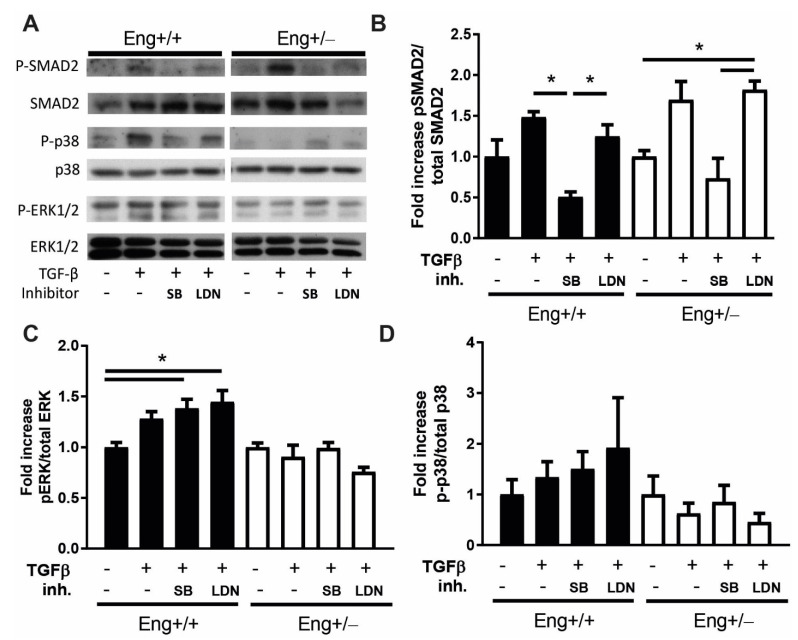
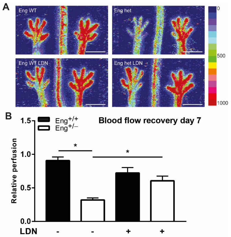
Hereditary hemorrhagic telangiectasia type 1 (HHT1) is a severe vascular disorder caused by mutations in the TGFβ/BMP co-receptor endoglin. Endoglin haploinsufficiency results in vascular malformations and impaired neoangiogenesis. Furthermore, HHT1 patients display an impaired immune response. To date it is not fully understood how endoglin haploinsufficient immune cells contribute to HHT1 pathology. Therefore, we investigated the immune response during tissue repair in Eng+/- mice, a model for HHT1. Eng+/- mice exhibited prolonged infiltration of macrophages after experimentally induced myocardial infarction. Moreover, there was an increased number of inflammatory M1-like macrophages (Ly6Chigh/CD206-) at the expense of reparative M2-like macrophages (Ly6Clow/CD206+). Interestingly, HHT1 patients also showed an increased number of inflammatory macrophages. In vitro analysis revealed that TGFβ-induced differentiation of Eng+/- monocytes into M2-like macrophages was blunted. Inhibiting BMP signaling by treating monocytes with LDN-193189 normalized their differentiation. Finally, LDN treatment improved heart function after MI and enhanced vascularization in both wild type and Eng+/- mice. The beneficial effect of LDN was also observed in the hind limb ischemia model. While blood flow recovery was hampered in vehicle-treated animals, LDN treatment improved tissue perfusion recovery in Eng+/- mice. In conclusion, BMPR kinase inhibition restored HHT1 macrophage imbalance in vitro and improved tissue repair after ischemic injury in Eng+/- mice.

Keywords: endoglin; hind limb ischemia; myocardial infarction; neovascularization; tissue repair; transforming growth factor-β.


Conflict of interest statement

The authors declare no conflict of interest.

Figures

Figure 1
Figure 1
Prolonged macrophage infiltration and decreased number of M2 macrophages after myocardial infarction in Eng+/−. (A) Cardiac sections of Eng+/+ and Eng+/− mice were stained for MAC-3 expressing macrophage (MAC-3 = brown; nuclei = blue) at day 4 and 14 post MI. Scale bar: 50 μm. (B) Quantification of the MAC3 positive cells shown in an N = 5–16 mice per group. (C) Splenic and cardiac tissue post-MI were stained for CD11b (general monocyte marker) and CD206 (M2 macrophage marker) 4 days after MI. Scale bar: 50 μm. (D) The ratio M1/M2 macrophages was determined by flow cytometry using a single cell suspension of Eng+/+ and Eng+/− mouse hearts 4 days post MI. The inflammatory M1 macrophage was identified by CD11b+/Ly6Chigh/CD206- selection and the regenerative M2 by CD11b+/Ly6Clow/CD206+ selection. N = 5–16 mice per group. Eng+/+ N = 9, Eng+/− N = 5. * p < 0.05, # p < 0.001, NS: not significant.
Figure 2
Figure 2
Macrophage phenotype is dependent on endoglin expression. (A) Macrophages isolated from Eng+/+ mice were stained with endoglin (red), Ly6C (green), and dapi (nuclei, blue). Scale bar: 10μm. (B) Representative flow charts of mouse and human isolated monocytes of Eng+/− mice and HHT1 patients and their healthy controls. Mouse inflammatory monocytes were distinguished by CD11b+/Ly6Chigh/CD206- and regenerative monocytes by CD11b+/Ly6Clow/CD206+ expression. Human inflammatory monocytes were distinguished by CD14+CD16- and regenerative monocytes by CD14+/CD16+ expression. (C) Quantification of the flow cytometry data as represented in B, divided in inflammatory and regenerative monocytes for mouse and human. Mouse samples: N = 5–16 mice per group. Eng+/+ N = 9, Eng+/− N = 5. Human samples: 7 HHT1 patients and 5 age- and gender-matched healthy human volunteers. * p < 0.05.
Figure 3
Figure 3
TGFβ signaling influences macrophage subtype differentiation. (A) Macrophages from Eng+/+ mice cultured with either GM-CSF for 7 days or in the presence of TGFβ (2.5 ng/µL) for 24 h and 96 h. The macrophage phenotype was determined based on the expression of Ly6C high (M1) and low (M2) of the CD11b expressing macrophages. * p = 0.001 difference in the number of M1 and M2 between GM-CSF vs. TGFβ stimulation for 96 h. (B,C) BM isolated monocytes from Eng+/+ (B) and Eng+/− (C) mice were cultured in the presence of GM-CSF in the presence or absence of TGFβ (2.5 ng/µL), SB (10 μM), or LDN (100 nM). The macrophage subtype was determined based on the expression of Ly6C high (M1) and low (M2) of the CD11b expressing macrophages. * p = 0.007; ** p < 0.0001.
Figure 4
Figure 4
Eng+/− macrophages show blunted TGFβ and BMP signaling responses in vitro. (A) Western blot analysis of Eng+/+ and Eng+/− cultured murine macrophages with GM-CSF, stimulated 60 min with TGFβ (2.5 ng/uL), SB (10 μM), and LDN (100 nM). Representative blots of N = 3 are shown. (B) Densitometric analysis of the blots shown in (A), expressed as percentage of phosphorylated Smad2 relative to total amount of Smad2. N = 3. (C) Quantification of the blots as shown in (A), expressed as percentage of phosphorylated ERK relative to total amount of ERK protein. N = 3. (D) Quantification of the blots in (A), expressed as the percentage of phosphorylated p38 relative to total amount of p38 protein, N = 3–4. Error bars are SEM. * p < 0.05.
Figure 5
Figure 5
LDN decreases p-Smad1 and induces p-Smad2 in the infarct border zone. Paraffin sections were stained for (A) pSmad1 or (C) pSmad2 and quantified in (B,D) for positive stained nuclei in Eng+/+ and Eng+/− mice treated with LDN or placebo. Representative images of heart sections 14 days post-MI are shown. N = 5–16 mice per group. Eng+/+ control N = 9, Eng+/− control N = 5, Eng+/+ LDN N = 6, Eng+/− LDN N = 16. Scale bars: 30 μm. * p < 0.05; # p < 0.001.
Figure 6
Figure 6
LDN restores cardiac function in Eng+/− to normal levels 14 days after MI. (A) Cardiac ejection fraction of Eng+/+ and Eng+/− mice 14 days post-MI, treated with LDN or placebo. N = 5–16 mice. (B) Infarct size was determined in both Eng+/+ and Eng+/− mice using Picrosirius Red staining. Top row—representative pictures of murine transversal heart sections. 1.0× magnification. Bottom row—quantification of infarcted area as percentage of total LV area. N = 5–16 mice per group. (C,D) LDN treatment influences cardiac neo-vascularization post-MI. (C) Paraffin sections of mouse hearts were stained for PECAM (green) and αSMA (red). N = 5–16 mice per group. Eng+/+ control N = 9, Eng+/− control N = 5, Eng+/+ LDN N = 6, Eng+/− LDN N = 16. (D,E) Quantification of the number of capillaries (D) and arteries (E) in (C). Scale bar: 50 μm. * p < 0.05; ** p < 0.01; # p < 0.001.
Figure 7
Figure 7
Hind limb blood flow recovery in female mice increases with LDN treatment. (A) Representative images of blood flow recovery in the paws as measured by laser Doppler perfusion imaging (LDPI), 7 days after HLI and subsequent treatment with LDN. Colors indicate the level of flow as indicated on the right panel of the figure. The left limb has HLI, the right limb was used as control. Scale bar: 1 cm (B) Quantification of LDPI measurements, N = 5–7 female mice per group. Black bars = WT, white bars = Eng+/−. * p < 0.05.

Similar articles

Cited by

References

    1. Guilhem A., Malcus C., Clarivet B., Plauchu H. Immunological abnormalities associated with hereditary haemorrhagic telangiec-tasia. J. Intern. Med. 2013:351–362. doi: 10.1111/joim.12098. - DOI - PubMed
    1. Pérez-Gómez E., Jerkic M., Prieto M., Del Castillo G., Martín-Villar E., Letarte M., Bernabeu C., Pérez-Barriocanal F., Quintanilla M., López-Novoa J.M. Impaired wound repair in adult endoglin heterozygous mice associated with lower NO bioavailability. J. Investig. Dermatol. 2014;134:247–255. doi: 10.1038/jid.2013.263. - DOI - PubMed
    1. Peter M.R., Jerkic M., Sotov V., Douda D.N., Ardelean D.S., Ghamami N., Lakschevitz F., Khan M.A., Robertson S.J., Glogauer M., et al. Impaired resolution of inflammation in the endoglin heterozygous mouse model of chronic colitis. Mediat. Inflamm. 2014;2014:1–13. doi: 10.1155/2014/767185. - DOI - PMC - PubMed
    1. Seghers L., De Vries M.R., Pardali E., Hoefer I.E., Hierck B.P., Dijke P.T.T., Goumans M.J., Quax P.H.A. Shear induced collateral artery growth modulated by endoglin but not by ALK1. J. Cell. Mol. Med. 2012;16:2440–2450. doi: 10.1111/j.1582-4934.2012.01561.x. - DOI - PMC - PubMed
    1. van Laake L.W., van den Driesche S., Post S., Feijen A., Jansen M.A., Driessens M.H., Mager J.J., Snijder R.J., Westermann C.J.J., Doevendans P.A., et al. Endoglin has a crucial role in blood cell-mediated vascular repair. Circulation. 2006;114:2288–2297. doi: 10.1161/CIRCULATIONAHA.106.639161. - DOI - PubMed

MeSH terms

LinkOut - more resources