. 2021 Feb 18;10(2):430.
doi: 10.3390/cells10020430.

Knockdown of AKT3 Activates HER2 and DDR Kinases in Bone-Seeking Breast Cancer Cells, Promotes Metastasis In Vivo and Attenuates the TGFβ/CTGF Axis

Affiliations

Knockdown of AKT3 Activates HER2 and DDR Kinases in Bone-Seeking Breast Cancer Cells, Promotes Metastasis In Vivo and Attenuates the TGFβ/CTGF Axis

Nico Hinz et al. Cells. .

Abstract

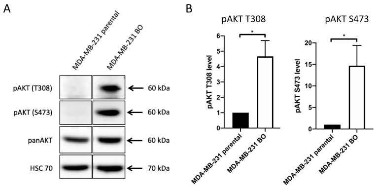
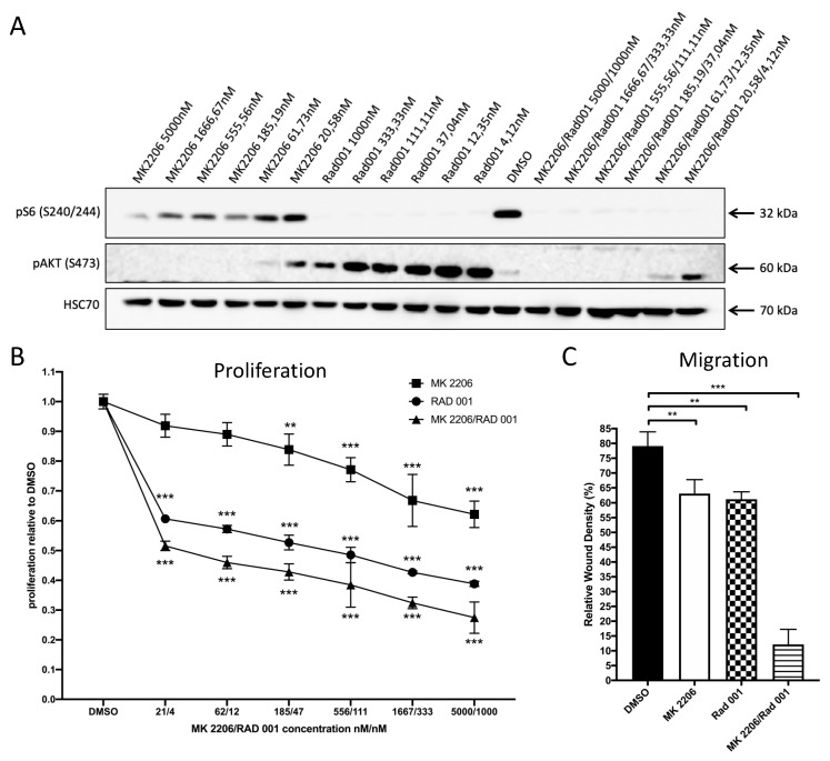
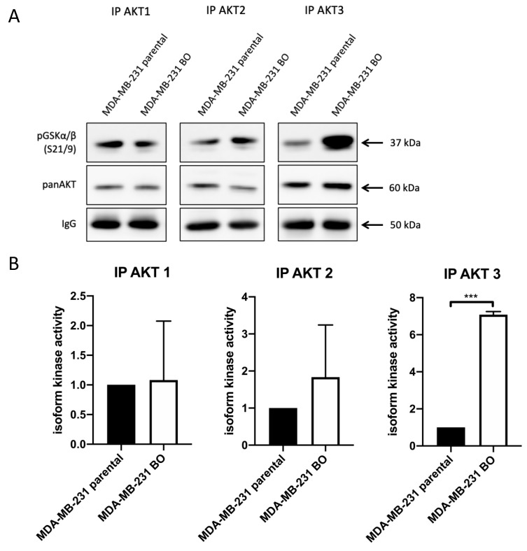
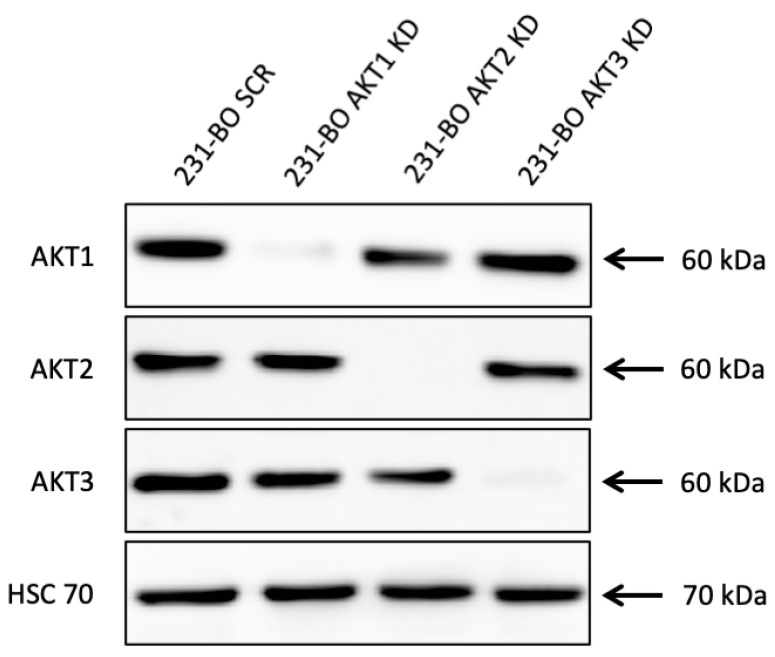
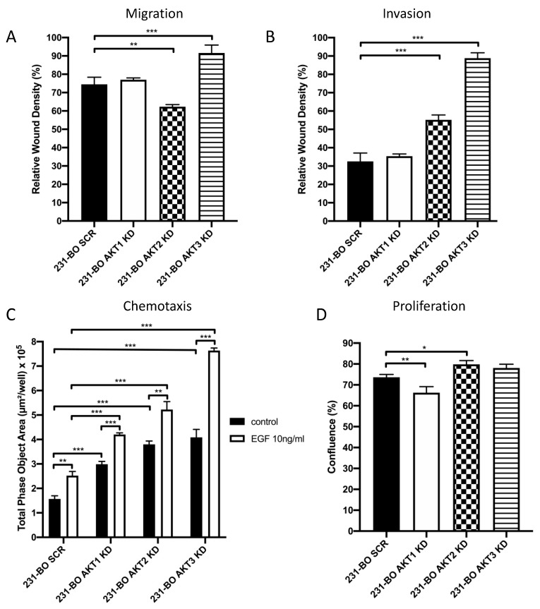
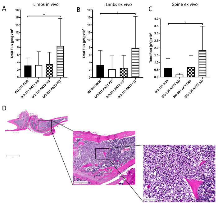
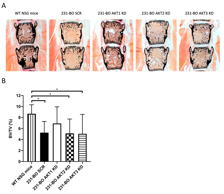
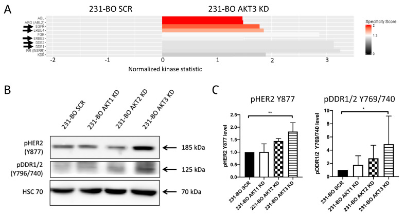
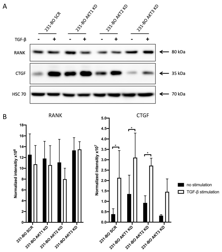
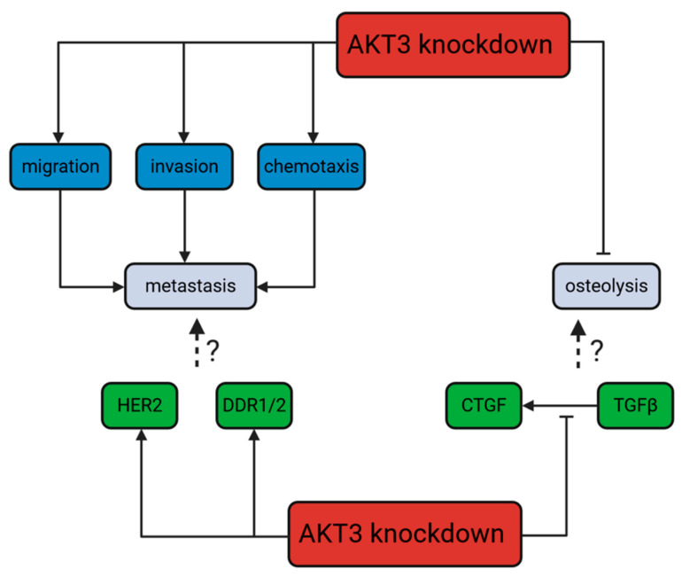
Bone metastases frequently occur in breast cancer patients and lack appropriate treatment options. Hence, understanding the molecular mechanisms involved in the multistep process of breast cancer bone metastasis and tumor-induced osteolysis is of paramount interest. The serine/threonine kinase AKT plays a crucial role in breast cancer bone metastasis but the effect of individual AKT isoforms remains unclear. Therefore, AKT isoform-specific knockdowns were generated on the bone-seeking MDA-MB-231 BO subline and the effect on proliferation, migration, invasion, and chemotaxis was analyzed by live-cell imaging. Kinome profiling and Western blot analysis of the TGFβ/CTGF axis were conducted and metastasis was evaluated by intracardiac inoculation of tumor cells into NOD scid gamma (NSG) mice. MDA-MB-231 BO cells exhibited an elevated AKT3 kinase activity in vitro and responded to combined treatment with AKT- and mTOR-inhibitors. Knockdown of AKT3 significantly increased migration, invasion, and chemotaxis in vitro and metastasis to bone but did not significantly enhance osteolysis. Furthermore, knockdown of AKT3 increased the activity and phosphorylation of pro-metastatic HER2 and DDR1/2 but lowered protein levels of CTGF after TGFβ-stimulation, an axis involved in tumor-induced osteolysis. We demonstrated that AKT3 plays a crucial role in bone-seeking breast cancer cells by promoting metastatic potential without facilitating tumor-induced osteolysis.

Keywords: AKT; AKT isoforms; bone metastasis; breast cancer; metastasis; organ tropism; osteolysis; vicious cycle.


Conflict of interest statement

The authors declare no conflict of interest.

Figures

Figure 1
Figure 1
The bone-seeking subline MDA-MB-231 BO exhibits constitutively elevated pAKT levels compared to MDA-MB-231 parental cells: (A) Western blot assay was performed to determine pAKT T308, pAKT S473 and panAKT levels using antibodies directed against the indicated proteins. HSC70 functions as a loading control. (B) Relative levels of pAKT T308 and pAKT S473 in the bone-seeking MDA-MB-231 subline compared to MDA-MB-231 parental cells were quantified from Western blot analysis of triplicates. Data were normalized to Ponceau staining and panAKT level. Bars indicate mean with SD; * = p < 0.05.
Figure 2
Figure 2
Treatment of 231-BO cells with the panAKT inhibitor MK2206 and the mTOR inhibitor RAD001 either alone or in combination inhibits proliferation and migration: (A) 231-BO cells were treated with MK2206 and RAD001 either alone or in combination with various concentrations for 24 h before preparing whole cell lysates. Inhibition of AKT (pAKT S473) and mTOR (pS6 S240/244) were confirmed by Western blot analysis using antibodies directed against the indicated proteins. HSC70 functions as a loading control. (B) 231-BO cells were seeded into a 96-well plate and were treated with various concentrations of MK2206 and RAD001 either alone or in combination for 72 h. Control cells were treated with DMSO. Cell viability was analyzed in triplicate using Alamar blue assay. Data points represent mean with SD; ** = p < 0.01; *** = p < 0.001. (C) Migration of 231-BO cells treated with MK2206 5000nM and RAD001 1000nM either alone or in combination was tested by scratch wound healing assay. Relative wound density was analyzed using the IncuCyte live cell imaging system. Bars indicate mean of triplicates with SD; ** = p < 0.01; *** = p < 0.001.
Figure 3
Figure 3
AKT3 activity is elevated in bone-seeking 231-BO cells in an in vitro kinase assay: (A) Isoform-specific in vitro AKT kinase assay was performed to determine differences in the bone-seeking subline 231-BO compared to parental MDA-MB-231 cells. After AKT isoform immunoprecipitation, GSK3α/β fusion protein was used as an AKT substrate. Level of pGSK3α/β S21/9 was detected as an indicator of AKT isoform activity. Antibodies directed against pGSK3α/β S21/9, panAKT, and mouse IgG were used after Western blotting. (B) Quantification of AKT isoform kinase activity indicated by phosphorylation of pGSK3α/β was performed from Western blot analysis of triplicates and normalized to intensity of IgG signals. Bars indicate mean relative to MDA-MB-231 parental cells with SD; *** = p < 0.001.
Figure 4
Figure 4
Lentiviral shRNA knockdown of AKT isoforms in 231-BO cells: Knockdowns of AKT isoforms were generated using lentiviral transduction of isoform-specific shRNA. Knockdown was confirmed by Western blot analysis and AKT1, AKT2, and AKT3 were detected with isoform-specific antibodies. HSC70 functions as a loading control.
Figure 5
Figure 5
Knockdown of AKT3 in 231-BO cells promotes migration, invasion, and chemotaxis towards EGF but has no effect on proliferation: (A) Migration of 231-BO cells with AKT isoform knockdowns was tested using a wound healing scratch assay. Relative wound density was analyzed from triplicates by the IncuCyte live cell imaging system. Bars indicate mean with SD; ** = p < 0.01, *** = p < 0.001. (B) 231-BO AKT isoform knockdown cells were seeded in a 96 well plate in triplicate for invasion assay. Matrigel was added as an ECM after scratch wounds were created. The IncuCyte live cell imaging system was used to determine relative wound density. Bars indicate mean with SD; *** = p < 0.001. (C) Chemotaxis of 231-BO cells with AKT isoform knockdowns was tested with a Boyden chamber assay using the IncuCyte live cell imaging system. Cells were seeded in the upper chamber and EGF in a concentration of 10ng/mL was added to the lower chamber. Boyden chambers without an EGF gradient served as control. The area of the lower chamber occupied with transmigrated cells through pores was determined. Bars indicate mean with SD; ** = p < 0.01, *** = p < 0.001. (D) 231-BO cells harboring AKT isoform knockdowns were seeded in a 96-well plate and confluence was monitored with the IncuCyte live cell imaging system. Bars indicate mean with SD; * = p < 0.05, ** = p < 0.01.
Figure 6
Figure 6
Knockdown of AKT3 in 231-BO cells shows increased metastasis to bone after intracardiac inoculation in NOD scid gamma (NSG) mice: To test the effect of AKT isoform knockdowns on metastasis in an in vivo model, 231-BO cells with AKT isoform knockdowns were transduced with a luciferase vector and were inoculated into the left ventricle of NSG mice. Correct injection, tumor cell dissemination, and growth were monitored by measurement of bioluminescence. Mice were sacrificed after 21 days, exenterated, and hind limbs as well as lumbar vertebrae were harvested. Bioluminescence intensities of hind limbs of living mice (SCR and AKT1 KD: n = 16; AKT2 KD and AKT3 KD n = 18) (A), of hind limbs ex vivo (SCR and AKT1 KD: n = 16; AKT2 KD and AKT3 KD n = 18) (B), and of the spine ex vivo (SCR and AKT1 KD: n = 8; AKT2 KD and AKT3 KD n = 9) (C) were measured by an IVIS imaging system after intraperitoneal luciferin injection. Bars indicate mean with SD; * = p < 0.05, ** = p < 0.01. (D) Right hind limbs were decalcified, embedded, and histological slices were generated. HE staining was used to confirm the presence of intraosseous tumor colonization. Image represents slices from a mouse injected with 231-BO AKT3 KD cells. Scale bars represent following dimensions: left image: 2000 μm; middle image: 500 μm; right image: 100 μm.
Figure 7
Figure 7
Knockdown of AKT3 in 231-BO cells does not enhance osteolysis in lumbar vertebral bodies after intracardiac injection: (A) Lumbar vertebrae bodies 1 to 5 were harvested from sacrificed mice after intracardiac injection of 231-BO cells with AKT isoform knockdowns and embedded in methylmetacrylate. Slices were stained with von Kossa/van Gieson staining to determine calcified bone volume. (B) Calcified bone volume was quantified relative to total tissue volume. WT NSG mice without tumor cell injection serve as a control (SCR, AKT1 KD, AKT2 KD, and AKT3 KD n = 25; WT NSG mice n = 6). Bars indicate mean with SD; * = p < 0.05.
Figure 8
Figure 8
Knockdown of AKT3 in 231-BO cells increases activity and phosphorylation of HER2 and DDR1/2: (A) To identify alterations in tyrosine kinase activity in the AKT3 knockdown of 231-BO cells, we performed functional kinome profiling of whole cell lysates. The resulting upstream kinase analysis of 231-BO SCR versus 231-BO AKT3 KD is depicted (normalized kinase statistic (log2) > 0: higher kinase activity in KD; specificity score (log2) > 1.3; white to red bars: statistically significant changes). Arrows indicate kinases that are discussed in the following. (B) Western blot analysis was conducted as a proof of principle for some identified upregulated kinases from kinome profiling. Antibodies directed against the indicated proteins were used. HSC70 functions as a loading control. (C) Relative levels of pHER2 Y877 and pDDR1/2 Y769/740 in 231-BO AKT isoform knockdowns compared to 231-BO SCR cells were quantified from Western blot analysis of triplicates. Data were normalized to Ponceau staining. Bars indicate mean with SD; * = p < 0.05; ** = p < 0.01.
Figure 9
Figure 9
Knockdown of AKT3 in 231-BO cells results in a diminished increase in CTGF expression after TGFβ-stimulation: (A) 231-BO cells with AKT isoform knockdowns were stimulated with 5ng/mL TGFβ for 24 h prior to cell lysate preparation. Western blot analysis of vicious cycle associated proteins was performed using antibodies directed against the indicated proteins. HSC70 functions as a loading control. (B) Expression of CTGF and RANK indicated by the normalized intensity in the 231-BO AKT isoform knockdowns after TGFβ-stimulation or without stimulation was quantified from Western blot analysis in triplicate. Data were normalized to Ponceau staining. Bars indicate mean with SD; * = p < 0.05.
Figure 10
Figure 10
Dichotomic role of an AKT3 knockdown in 231-BO cells: promotion of metastasis but not osteolysis: On the one hand, knockdown of AKT3 increases, among others, DDR1/2 and HER2 signaling in bone-seeking breast cancer cells as well as promoting migration, invasion, and chemotaxis as steps of the metastatic process in general. HER2 and DDR1/2 activity is associated with metastasis in breast cancer in the literature. On the other hand, knockdown of AKT3 decreases the TGFβ-stimulated CTGF expression. In connection to these findings, knockdown of AKT3 leads to an absent promotion of osteolysis. The TGFβ/CTGF axis is correlated with the vicious cycle of osteolysis in breast cancer in the literature.

Similar articles

Cited by

References

    1. Siegel R.L., Miller K.D., Jemal A. Cancer statistics, 2019. CA Cancer J. Clin. 2019;69:7–34. doi: 10.3322/caac.21551. - DOI - PubMed
    1. Berman A.T., Thukral A.D., Hwang W.T., Solin L.J., Vapiwala N. Incidence and patterns of distant metastases for patients with early-stage breast cancer after breast conservation treatment. Clin. Breast Cancer. 2013;13:88–94. doi: 10.1016/j.clbc.2012.11.001. - DOI - PubMed
    1. Mundy G.R. Metastasis to bone: Causes, consequences and therapeutic opportunities. Nat. Rev. Cancer. 2002;2:584–593. doi: 10.1038/nrc867. - DOI - PubMed
    1. Hiraga T., Williams P.J., Mundy G.R., Yoneda T. The bisphosphonate ibandronate promotes apoptosis in MDA-MB-231 human breast cancer cells in bone metastases. Cancer Res. 2001;61:4418–4424. - PubMed
    1. Goldhirsch A., Wood W.C., Coates A.S., Gelber R.D., Thurlimann B., Senn H.J., Panel m. Strategies for subtypes--dealing with the diversity of breast cancer: Highlights of the St. Gallen International Expert Consensus on the Primary Therapy of Early Breast Cancer 2011. Ann. Oncol. 2011;22:1736–1747. doi: 10.1093/annonc/mdr304. - DOI - PMC - PubMed

Publication types

MeSH terms