

A Facile Strategy for Fabrication Lysozyme-Loaded Mesoporous Silica Nanotubes from Electrospun Silk Fibroin Nanofiber Templates
- PMID: 33670610
- PMCID: PMC7923156
- DOI: 10.3390/molecules26041073
A Facile Strategy for Fabrication Lysozyme-Loaded Mesoporous Silica Nanotubes from Electrospun Silk Fibroin Nanofiber Templates
Abstract
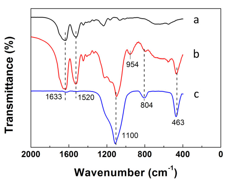
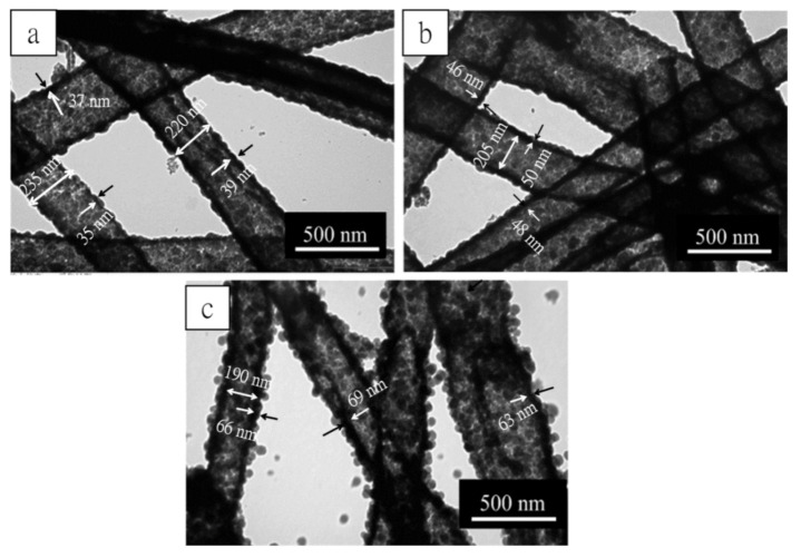
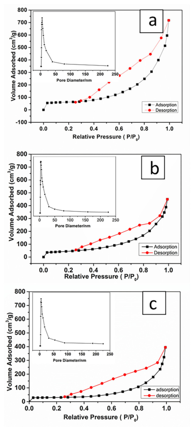
This paper presents a facile and low-cost strategy for fabrication lysozyme-loaded mesoporous silica nanotubes (MSNTs) by using silk fibroin (SF) nanofiber templates. The "top-down method" was adopted to dissolve degummed silk in CaCl2/ formic acid (FA) solvent, and the solution containing SF nanofibrils was used for electrospinning to prepare SF nanofiber templates. As SF contains a large number of -OH, -NH2 and -COOH groups, the silica layer could be easily formed on its surface by the Söber sol-gel method without adding any surfactant or coupling agent. After calcination, the MSNTs were obtained with inner diameters about 200 nm, the wall thickness ranges from 37 ± 2 nm to 66 ± 3 nm and the Brunauer-Emmett-Teller (BET) specific surface area was up to 200.48 m2/g, the pore volume was 1.109 cm3/g. By loading lysozyme, the MSNTs exhibited relatively high drug encapsulation efficiency up to 31.82% and an excellent long-term sustained release in 360 h (15 days). These results suggest that the MSNTs with the hierarchical structure of mesoporous and macroporous will be a promising carrier for applications in biomacromolecular drug delivery systems.
Keywords: drug release; electrospinning; lysozyme; mesoporous silica nanotubes; silk fibroin.
Conflict of interest statement
The authors declare no conflict of interest.
Figures


Similar articles
-
Electrospun nanofibers comprising of silk fibroin/gelatin for drug delivery applications: Thyme essential oil and doxycycline monohydrate release study.J Biomed Mater Res A. 2018 Apr;106(4):1092-1103. doi: 10.1002/jbm.a.36303. Epub 2018 Jan 3. J Biomed Mater Res A. 2018. PMID: 29210169
-
Fluorescent mesoporous silica nanotubes incorporating CdS quantum dots for controlled release of ibuprofen.Acta Biomater. 2009 Nov;5(9):3488-96. doi: 10.1016/j.actbio.2009.05.002. Epub 2009 May 13. Acta Biomater. 2009. PMID: 19442764
-
Development of artificial dermis using 3D electrospun silk fibroin nanofiber matrix.J Biomed Nanotechnol. 2014 Jul;10(7):1294-303. doi: 10.1166/jbn.2014.1818. J Biomed Nanotechnol. 2014. PMID: 24804550
-
Silk industry waste protein: isolation, purification and fabrication of electrospun silk protein nanofibers as a possible nanocarrier for floating drug delivery.Nanotechnology. 2021 Jan 15;32(3):035101. doi: 10.1088/1361-6528/abb8a9. Nanotechnology. 2021. PMID: 32932237
-
Functionalized silk fibroin nanofibers as drug carriers: Advantages and challenges.J Control Release. 2020 May 10;321:324-347. doi: 10.1016/j.jconrel.2020.02.022. Epub 2020 Feb 13. J Control Release. 2020. PMID: 32061791 Review.
Cited by
-
Starch-Directed Synthesis of Worm-Shaped Silica Microtubes.Materials (Basel). 2023 Apr 2;16(7):2831. doi: 10.3390/ma16072831. Materials (Basel). 2023. PMID: 37049125 Free PMC article.
-
Advances in Preparation and Properties of Regenerated Silk Fibroin.Int J Mol Sci. 2023 Aug 24;24(17):13153. doi: 10.3390/ijms241713153. Int J Mol Sci. 2023. PMID: 37685960 Free PMC article. Review.
-
Release Profiles of Carvacrol or Chlorhexidine of PLA/Graphene Nanoplatelets Membranes Prepared Using Electrospinning and Solution Blow Spinning: A Comparative Study.Molecules. 2023 Feb 19;28(4):1967. doi: 10.3390/molecules28041967. Molecules. 2023. PMID: 36838955 Free PMC article.
References
-
- Bao Y., Shi C., Wang T., Li X., Ma J. Recent progress in hollow silica: Template synthesis, morphologies and applications. Microporous Mesoporous Mater. 2016;227:121–136. doi: 10.1016/j.micromeso.2016.02.040. - DOI
-
- Wu C., Yuan W., Al-Deyab S.S., Zhang K.-Q. Tuning porous silica nanofibers by colloid electrospinning for dye adsorption. Appl. Surf. Sci. 2014;313:389–395. doi: 10.1016/j.apsusc.2014.06.002. - DOI
-
- Peng Y., Leng W., Dong B., Ge R., Duan H., Gao Y. Bottom-up preparation of gold nanoparticle-mesoporous silica composite nanotubes as a catalyst for the reduction of 4-nitrophenol. Chin. J. Catal. 2015;36:1117–1123. doi: 10.1016/S1872-2067(14)60310-7. - DOI
-
- Yang X., He D., He X., Wang K., Tang J., Zou Z., He X., Xiong J., Li L., Shangguan J. Synthesis of Hollow Mesoporous Silica Nanorods with Controllable Aspect Ratios for Intracellular Triggered Drug Release in Cancer Cells. ACS Appl. Mater. Interfaces. 2016;8:20558–20569. doi: 10.1021/acsami.6b05065. - DOI - PubMed
MeSH terms
Substances
Grants and funding
LinkOut - more resources
Full Text Sources
Other Literature Sources
Research Materials
