Review
. 2021 Feb 18;22(4):2022.
doi: 10.3390/ijms22042022.

The Neurovascular Unit Dysfunction in Alzheimer's Disease

Affiliations
Review

The Neurovascular Unit Dysfunction in Alzheimer's Disease

Luis O Soto-Rojas et al. Int J Mol Sci. .

Abstract

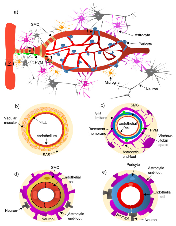
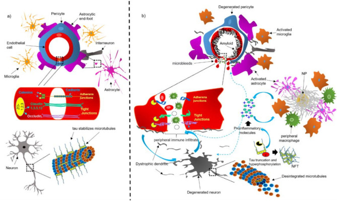
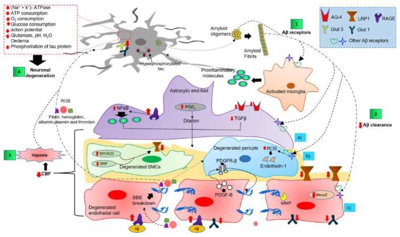
Alzheimer's disease (AD) is the most common neurodegenerative disease worldwide. Histopathologically, AD presents with two hallmarks: neurofibrillary tangles (NFTs), and aggregates of amyloid β peptide (Aβ) both in the brain parenchyma as neuritic plaques, and around blood vessels as cerebral amyloid angiopathy (CAA). According to the vascular hypothesis of AD, vascular risk factors can result in dysregulation of the neurovascular unit (NVU) and hypoxia. Hypoxia may reduce Aβ clearance from the brain and increase its production, leading to both parenchymal and vascular accumulation of Aβ. An increase in Aβ amplifies neuronal dysfunction, NFT formation, and accelerates neurodegeneration, resulting in dementia. In recent decades, therapeutic approaches have attempted to decrease the levels of abnormal Aβ or tau levels in the AD brain. However, several of these approaches have either been associated with an inappropriate immune response triggering inflammation, or have failed to improve cognition. Here, we review the pathogenesis and potential therapeutic targets associated with dysfunction of the NVU in AD.

Keywords: Alzheimer’s disease; amyloid peptide; astrocytes; blood-brain barrier; microglia; tau protein.


Conflict of interest statement

The authors declare no conflict of interest.

Figures

Figure 1
Figure 1
Illustration of the constitutive cellular elements of the neurovascular unit (NVU) along the entire brain vasculature. Panel (a) shows a panoramic view of the vascular tree. Panels (be) depict the different levels of the vascular tree. Pial arteries (panel (b)) run along the brain surface and penetrate the parenchyma, narrowing and branching into penetrating arterioles (panel (c)), which form the intraparenchymal arterioles (panel (d)), and ultimately give rise to capillaries (panel (e)). Abbreviations: IEL, internal elastic lamina; PVM, perivascular macrophage; SAS, the subarachnoid space; SMC, smooth muscle cell.
Figure 2
Figure 2
Schematic representation of the NVU under physiological (a) and pathological (b) conditions. Abbreviations: NFT, neurofibrillary tangle; NP, neuritic plaque.
Figure 3
Figure 3
Overview of the complex cell–cell signaling in the NVU, triggered by extracellular and vascular Aβ deposits. Step 1 shows the interaction of Aβ oligomers and fibrils with NVU cells through several receptors. Step 2 illustrates the altered Aβ clearance that triggers NVU dysfunction. Step 3 shows the NVU cell dysfunction as the causative agent of cerebral hypoxia. Step 4 shows cerebral hypoxia, neuroinflammatory environment, and peripheral blood infiltrate as the promoting causes of neuronal degeneration. Abbreviations: AQ-4, aquaporin-4; Aβ, amyloid peptide; BBB, blood-brain barrier; CBF, cerebral blood flow; GLUT, glucose transporter; LRP1, low-density lipoprotein receptor-related protein 1; MEOX2, mesenchyme homeobox 2; MMP, matrix metalloproteinase; MYOCD, myocardin; NFκB, nuclear factor kappa light chain enhancer of activated B cells; PDGF-B, platelet-derived growth factor subunit B; PDGFRβ, platelet-derived growth factor receptor β; PGE2, prostaglandin E2; TGFβ, transforming growth factor-beta; RAGE, receptor for advanced glycation end products; ROS, reactive oxygen species.

References

    1. Harder D.R., Zhang C., Gebremedhin D. Astrocytes function in matching blood flow to metabolic activity. News Physiol. Sci. 2002;17:27–31. doi: 10.1152/physiologyonline.2002.17.1.27. - DOI - PubMed
    1. Zlokovic B.V. The blood-brain barrier in health and chronic neurodegenerative disorders. Neuron. 2008;57:178–201. doi: 10.1016/j.neuron.2008.01.003. - DOI - PubMed
    1. Abbott N.J., Friedman A. Overview and introduction: The blood-brain barrier in health and disease. Epilepsia. 2012;53:1–6. doi: 10.1111/j.1528-1167.2012.03696.x. - DOI - PMC - PubMed
    1. Armulik A., Genove G., Mae M., Nisancioglu M.H., Wallgard E., Niaudet C., He L., Norlin J., Lindblom P., Strittmatter K., et al. Pericytes regulate the blood-brain barrier. Nature. 2010;468:557–561. doi: 10.1038/nature09522. - DOI - PubMed
    1. Winkler E.A., Bell R.D., Zlokovic B.V. Central nervous system pericytes in health and disease. Nat. Neurosci. 2011;14:1398–1405. doi: 10.1038/nn.2946. - DOI - PMC - PubMed

LinkOut - more resources