. 2021 Feb 28;13(3):385.
doi: 10.3390/v13030385.

Isolation of a New Infectious Pancreatic Necrosis Virus (IPNV) Variant from a Fish Farm in Scotland

Affiliations

Isolation of a New Infectious Pancreatic Necrosis Virus (IPNV) Variant from a Fish Farm in Scotland

Jessica Benkaroun et al. Viruses. .

Abstract

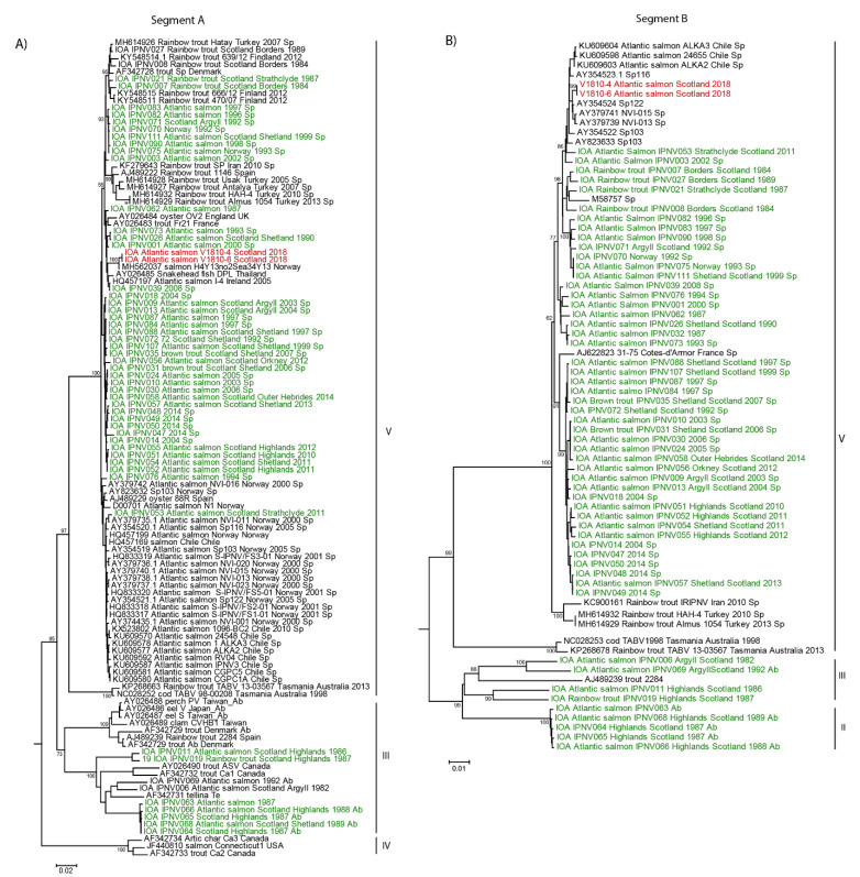
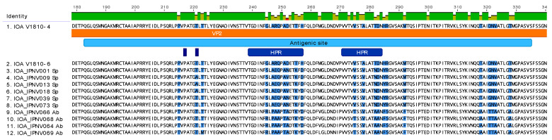
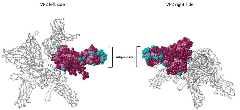
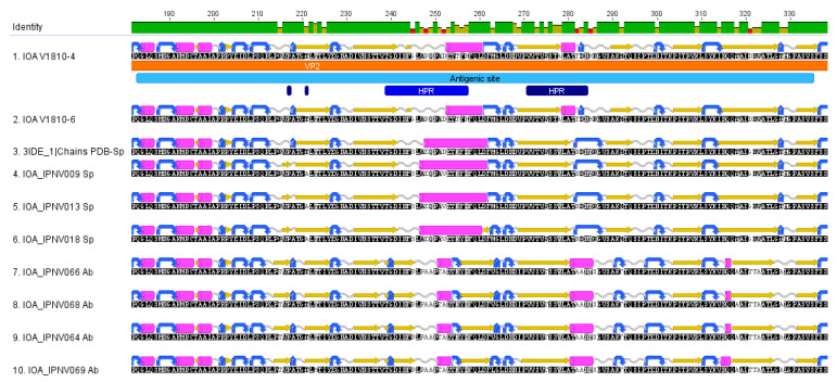
The aquatic virus, infectious pancreatic necrosis virus (IPNV), is known to infect various farmed fish, in particular salmonids, and is responsible for large economic losses in the aquaculture industry. Common practices to detect the virus include qPCR tests based on specific primers and serum neutralization tests for virus serotyping. Following the potential presence of IPNV viruses in a fish farm in Scotland containing vaccinated and IPNV-resistant fish, the common serotyping of the IPNV isolates was not made possible. This led us to determine the complete genome of the new IPNV isolates in order to investigate the cause of the serotyping discrepancy. Next-generation sequencing using the Illumina technology along with the sequence-independent single primer amplification (SISPA) approach was conducted to fully characterize the new Scottish isolates. With this approach, the full genome of two isolates, V1810-4 and V1810-6, was determined and analyzed. The potential origin of the virus isolates was investigated by phylogenetic analyses along with tridimensional and secondary protein structure analyses. These revealed the emergence of a new variant from one of the main virus serotypes, probably caused by the presence of selective pressure exerted by the vaccinated IPNV-resistant farmed fish.

Keywords: IPNV; aquaculture; evolution; infectious pancreatic necrosis virus; phylogeny; variant.


Conflict of interest statement

The authors declare no conflict of interests.

Figures

Figure 1
Figure 1
Phylogeny of the new Scottish infectious pancreatic necrosis virus (IPNV) isolates V1810. Nucleotide sequences of the new isolates from Scotland, V1810–4 and V1810–6, were aligned with previous Scottish isolates and similar sequences retrieved by BLAST on NCBI. The alignment was conducted by MUSCLE implemented in MEGA 6. The phylogenetic tree was inferred in MEGA6 with the neighbor-joining method. Bootstrap percentages >70 are indicated. (A) Phylogenetic tree of IPNV segment A. (B) Phylogenetic tree of IPNV segment B. Sequences on the trees colored in red correspond to the new Scottish isolates V1810–4 and V1810–6; in green are previous Scottish isolates, and in black are sequences retrieved by BLAST on NCBI.
Figure 2
Figure 2
Analysis of the VP2 protein sequences of the new Scottish IPNV isolates at the antigenic site. The new Scottish isolates V1810–4, and V1810–6 amino acid sequences were aligned with Scottish isolates of serotype Sp or Ab with MUSCLE. The sequence was annotated manually in Geneious Prime v.2021.0.3 [32]. Zoom representation of the VP2 protein at the antigenic site. The antigenic site is contained in the VP2 protein (colored in clear blue), HPR regions are colored in dark blue. Amino acid residues variation is highlighted in blue on the sequences.
Figure 3
Figure 3
3D structural analysis of the VP2 protein. The conservation of the amino acid residues was estimated using the ConSurf Server [33] and based on the multiple alignments of Sp and Ab serotypes along with the 3IDE_1 PDB 3D structure. The left and right view of the protein is represented. One VP2 monomer is colored on the 3D structure of VP2, which is composed of 4 monomers. Conserved amino acid residues are colored in purple. Pink residues, less conserved than purple residues, are colored in pink. The least conserved residues are colored in blue.
Figure 4
Figure 4
Secondary structures of VP2 proteins. The secondary structures of VP2 proteins among the Sp and Ab isolates and the new V1810–4 and V1810–6 isolates are depicted as follows: Beta sheets are colored in yellow, turns in blue, alpha-helices in pink, and coil structures in clear gray. The secondary structures were predicted with EMBOSS implemented in Geneious Prime. All amino acid sequences are oriented from the amine terminal extremity to the carboxy-terminal extremity.

Similar articles

Cited by

References

    1. Dobos P. The molecular biology of infectious pancreatic necrosis virus (IPNV) Annu. Rev. Fish Dis. 1995;5:25–54. doi: 10.1016/0959-8030(95)00003-8. - DOI
    1. Dobos P. Protein-primed RNA synthesis in vitro by the virion-associated RNA polymerase of infectious pancreatic necrosis virus. Virology. 1995;208:19–25. doi: 10.1006/viro.1995.1125. - DOI - PubMed
    1. Duncan R., Dobos P. The nucleotide sequence of infectious pancreatic necrosis virus (IPNV) dsRNA segment A reveals one large ORF encoding a precursor polyprotein. Nucleic Acids Res. 1986;14:5934. doi: 10.1093/nar/14.14.5934. - DOI - PMC - PubMed
    1. Duncan R., Nagy E., Krell P.J., Dobos P.E. Synthesis of the infectious pancreatic necrosis virus polyprotein, detection of a virus-encoded protease, and fine structure mapping of genome segment A coding regions. J. Virol. 1987;61:3655–3664. doi: 10.1128/JVI.61.12.3655-3664.1987. - DOI - PMC - PubMed
    1. Petit S., Lejal N., Huet J.C., Delmas B. Active residues and viral substrate cleavage sites of the protease of the birnavirus infectious pancreatic necrosis virus. J. Virol. 2000;74:2057–2066. doi: 10.1128/JVI.74.5.2057-2066.2000. - DOI - PMC - PubMed

Publication types

Substances

LinkOut - more resources