Review
. 2021 Feb 28;22(5):2434.
doi: 10.3390/ijms22052434.

An Extracellular Perspective on CNS Maturation: Perineuronal Nets and the Control of Plasticity

Affiliations
Review

An Extracellular Perspective on CNS Maturation: Perineuronal Nets and the Control of Plasticity

Daniela Carulli et al. Int J Mol Sci. .

Abstract

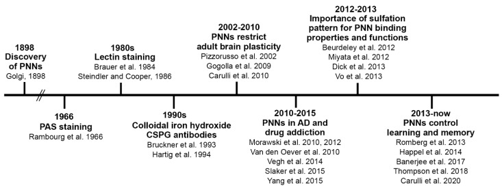
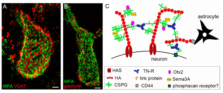
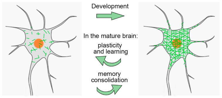
During restricted time windows of postnatal life, called critical periods, neural circuits are highly plastic and are shaped by environmental stimuli. In several mammalian brain areas, from the cerebral cortex to the hippocampus and amygdala, the closure of the critical period is dependent on the formation of perineuronal nets. Perineuronal nets are a condensed form of an extracellular matrix, which surrounds the soma and proximal dendrites of subsets of neurons, enwrapping synaptic terminals. Experimentally disrupting perineuronal nets in adult animals induces the reactivation of critical period plasticity, pointing to a role of the perineuronal net as a molecular brake on plasticity as the critical period closes. Interestingly, in the adult brain, the expression of perineuronal nets is remarkably dynamic, changing its plasticity-associated conditions, including memory processes. In this review, we aimed to address how perineuronal nets contribute to the maturation of brain circuits and the regulation of adult brain plasticity and memory processes in physiological and pathological conditions.

Keywords: Alzheimer’s disease; chondroitin sulfate proteoglycans; critical period; drug addiction; learning; memory; perineuronal net.


Conflict of interest statement

The authors declare no conflict of interest. The funders had no role in the design of the study; in the collection, analyses, or interpretation of data; in the writing of the manuscript, or in the decision to publish the results.

Figures

Figure 2
Figure 2
Timeline of key PNN-related discoveries. The timeline shows some of the critical contributions to the PNN field, from the first papers in which PNNs were visualized to the first studies elucidating PNN molecular composition and PNNs’ role in plasticity and disease. AD: Alzheimer’s disease, PAS: periodic acid–Schiff.
Figure 1
Figure 1
Structure and composition of the PNN. (A,B) show PNNs around neurons in the mouse cerebellar nuclei, labelled by Wisteria floribunda agglutinin (WFA), in green. PNNs display their typical holes, in which pre-synaptic terminals are contained. In (A), GABAergic terminals are shown (in red), labelled by anti-VGAT antibodies. In (B), post-synaptic clusters of gephyrin, which anchors GABA receptors to the underlying cytoskeleton, are shown (in red). In (C), the main molecular components of PNNs are depicted. Scale bar: 4 µm in (A,B).
Figure 3
Figure 3
Dynamics of PNNs. The scheme shows that PNN components accumulate and gather around the cell body, proximal dendrites and axon initial segment of a neuron during postnatal development. However, once formed, the PNN is not a static structure. In the adult brain PNNs are reduced, for instance, during learning, and restored when the learning phase is terminated and memories are consolidated.

Similar articles

Cited by

References

    1. Reh R.K., Dias B.G., Nelson C.A., 3rd, Kaufer D., Werker J.F., Kolb B., Levine J.D., Hensch T.K. Critical period regulation acrossmultiple timescales. Proc. Natl. Acad. Sci. USA. 2020;117:23242–23251. doi: 10.1073/pnas.1820836117. - DOI - PMC - PubMed
    1. Hubel D.H., Wiesel T.N. Receptive fields, binocular interaction and functional architecture in the cat’s visual cortex. J. Physiol. 1962;160:106–154. doi: 10.1113/jphysiol.1962.sp006837. - DOI - PMC - PubMed
    1. Hubel D.H., Wiesel T.N. Binocular interaction in striate cortex of kittens reared with artificial squint. J. Neurophysiol. 1965;28:1041–1059. doi: 10.1152/jn.1965.28.6.1041. - DOI - PubMed
    1. Fifková E. Changes in the visual cortex of rats after unilateral deprivation. Nature. 1968;220:379–381. doi: 10.1038/220379a0. - DOI - PubMed
    1. Antonini A., Stryker M.P. Plasticity of geniculocortical afferents following brief or prolonged monocular occlusion in the cat. J. Comp. Neurol. 1996;369:64–82. doi: 10.1002/(SICI)1096-9861(19960520)369:1<64::AID-CNE5>3.0.CO;2-I. - DOI - PubMed