Review
. 2021 Feb 28;13(3):387.
doi: 10.3390/v13030387.

From Submerged Cultures to 3D Cell Culture Models: Evolution of Nasal Epithelial Cells in Asthma Research and Virus Infection

Affiliations
Review

From Submerged Cultures to 3D Cell Culture Models: Evolution of Nasal Epithelial Cells in Asthma Research and Virus Infection

Malik Aydin et al. Viruses. .

Abstract

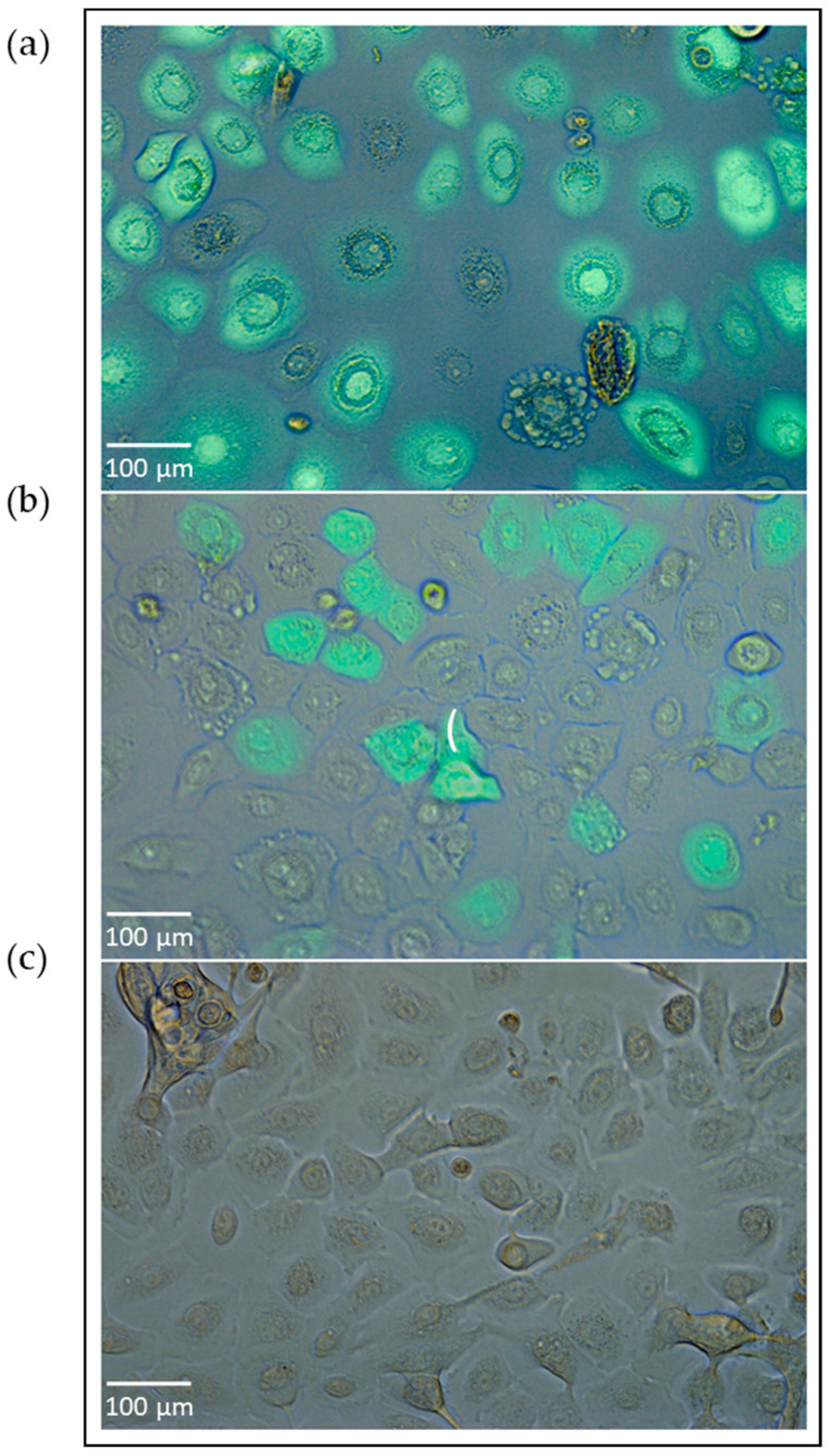
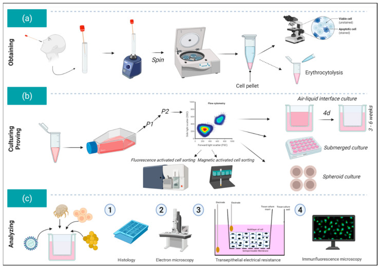
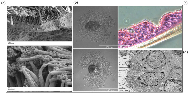
Understanding the response to viral infection in the context of respiratory diseases is of significant importance. Recently, there has been more focus on the role of the nasal epithelium in disease modeling. Here, we provide an overview of different submerged, organotypic 3D and spheroid cell culture models of nasal epithelial cells, which were used to study asthma and allergy with a special focus on virus infection. In detail, this review summarizes the importance, benefits, and disadvantages of patient-derived cell culture models of nasal- and bronchial epithelial cells, including a comparison of these cell culture models and a discussion on why investigators should consider using nasal epithelial cells in their research. Exposure experiments, simple virus transduction analyses as well as genetic studies can be performed in these models, which may provide first insights into the complexity of molecular signatures and may open new doors for drug discovery and biomarker research.

Keywords: 3D cell culture; air–liquid-interface; asthma; bronchial epithelial cells; culture techniques; epithelial cells; nasal epithelial cells; spheroids; submerged; virus.


Conflict of interest statement

The authors declare no conflict of interest. The funders had no role in the design of the study; in the collection, analyses, or interpretation of data; in the writing of the manuscript, or in the decision to publish the results. FP receives royalties from Elsevier for the 24th Ed. of the anatomy atlas “Sobotta” and the “Sobotta Textbook of Anatomy”. The German Research Foundation that supported FP (PA738/15-1) had no role in the design or conduct of this research.

Figures

Figure 1
Figure 1
The submerged culture of nasal epithelial cells transduced with adenovirus. The cultivation of nasal epithelial cells derived from a healthy subject from our pediatric exacerbation study cohort [96]. Nasal epithelial cells were seeded in an amount of 20,000 cells per well and resuspended in BEGMTM medium (Lonza, Basel, Switzerland) in collagen-coated 24-well culture plates prior to virus transduction. The next day, previously established recombinant adenovirus type 5 encoding green fluorescent protein [97] was added in different virus particle concentrations (vpc) (e.g., 1 × 10⁸ vpc), incubated for one day upon viral transduction, and the transduction efficiency was analyzed through immunofluorescence microscopy. The subfigures (a,b) represent virus transduction efficiencies in nasal epithelial cells directly correlating with virus transduction rates. The picture (c) shows the nasal epithelial cells without viral transduction (untreated, wildtype cells). This experiment was separately performed for this manuscript, correlates thematically with [22], and was not previously published elsewhere.
Figure 2
Figure 2
An overview of the workflow for analyzing primary human nasal epithelial cells. (a) After careful collection of the nasal epithelial cells from the nasal cavity using a cytology or interdental brush, cells are transferred to a 15 mL tube and shaken several times. The brush is then removed, and the cells are centrifuged. The cells are checked for viability using a Neubauer counting chamber using trypan blue or Türk’s solution (purchased from Sigma Aldrich). If necessary, erythrocytolysis can be also performed separately. (b) The cells are then seeded into collagen-coated T25/T75 flasks in, for example, BEGMTM medium. After two passages, flow cytometric characterization of the cells can be performed if needed. If the cultures lack purity, separation by Magnetic-activated cell sorting (MACS) or Fluorescence-activated cell sorting (FACS) can be also performed (usually these steps are not necessary). Afterwards the nasal epithelial cells are either transferred into air–liquid interface cultures (ALI) (approximately 9–11 × 104 cells per 1.12 cm2 or 2–3 × 10⁴ cells per 0.33 cm2 cell culture inserts = transwells), or directly seeded in submerged cultures. For certain questions, nasal epithelial cells can be also seeded for spheroid cultures (circa 1000 cells per U-bottom well). (c) Different provocation experiments or co-cultures can be performed with the cultures (here schematic presentation for ALI cultures) and subsequently, the morphology of the cells can be studied through histology and electron microscopy [22]. By means of transepithelial electrical resistance measurement, the integrity of the cell–cell interaction can be measured, or the permissiveness can be investigated through immunofluorescence microscopic analysis. This content of this figure correlates thematically with [22] and was not previously published elsewhere. This figure was created with www.biorender.com (accessed on 27 February 2021).
Figure 3
Figure 3
The morphology of nasal epithelial cells. (a) Scanning electron microscopic imaging of organotypic 3D nasal epithelial cell cultures derived from young children with asthma or healthy subjects (left top: ×4.00 K; left bottom: ×9.45K, in different sizes). These images show the morphology of the nasal epithelial cell culture with cilia, the integrity of the cell–cell-interaction, and the glycocalyx structure. The samples were prepared for imaging with Zeiss Sigma VP SEM (Zeiss, Oberkochen, Germany) scanning electron microscope at 2 kV acceleration voltage using the in-lens and SE detectors. (b) Nasal epithelial cells were seeded for the submerged spheroid culture technique. Cells derived from a healthy subject were seeded in a 96-well plate at circa 1000 cells per well in U-Bottom plate (×10 magnification, size 200 µm) receiving PneumaCultTM ALI medium (STEMCELLTM Technologies, Vancouver, Canada). After reaching a determined cell size, the spheroid culture can be used for distinct experimental questions. This experiment was separately performed and correlates thematically with [22]. (c) ALI culture derived from a healthy subject. After passaging nasal epithelial cells from the 2D submerged into the organotypic 3D ALI culture model, samples were prepared for histological analyses. This figure presents the cilia morphology of the cultures and the cell–cell integrity in PAS reaction. The ciliary epithelium is precisely imaged (×10 magnification, size 50 µm). (d) Transmission electron microscopic (TEM) analysis of nasal epithelial ALI cultures (×20,000 magnification, size 2.500 µm). The aim of this additional method and figure is to present the cell–cell–contacts (tight junctions), cilia and organelles through TEM to study the effectiveness of the cell culture model. These methods/pictures were separately performed, correlate thematically with [22] and were not previously published elsewhere.

Similar articles

Cited by

References

    1. Braman S.S. The global burden of asthma. Chest. 2006;130:4S–12S. doi: 10.1378/chest.130.1_suppl.4S. - DOI - PubMed
    1. Lambrecht B.N., Hammad H. The immunology of asthma. Nat. Immunol. 2015;16:45–56. doi: 10.1038/ni.3049. - DOI - PubMed
    1. Asher I., Pearce N. Global burden of asthma among children. Int. J. Tuberc Lung Dis. 2014;18:1269–1278. doi: 10.5588/ijtld.14.0170. - DOI - PubMed
    1. Lambrecht B.N., Hammad H. The airway epithelium in asthma. Nat. Med. 2012;18:684–692. doi: 10.1038/nm.2737. - DOI - PubMed
    1. Wenzel S.E. Asthma phenotypes: The evolution from clinical to molecular approaches. Nat. Med. 2012;18:716–725. doi: 10.1038/nm.2678. - DOI - PubMed

Publication types