. 2021 Feb 28;10(3):366.
doi: 10.3390/antiox10030366.

Protective Effect of Oligonol on Dimethylnitrosamine-Induced Liver Fibrosis in Rats via the JNK/NF-κB and PI3K/Akt/Nrf2 Signaling Pathways

Affiliations

Protective Effect of Oligonol on Dimethylnitrosamine-Induced Liver Fibrosis in Rats via the JNK/NF-κB and PI3K/Akt/Nrf2 Signaling Pathways

Changyong Lee et al. Antioxidants (Basel). .

Abstract

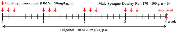
Oligonol is a low molecular weight polyphenol product derived from lychee fruit by a manufacturing process. We investigated oligonol's anti-fibrotic effect and the underlying mechanism in dimethylnitrosamine (DMN)-induced chronic liver damage in male Sprague-Dawley rats. Oral administration of oligonol (10 and 20 mg/kg body weight) ameliorated the DMN-induced abnormalities in liver histology and serum parameters in rats. Oligonol prevented the DMN-induced elevations of TNF-α, IL-1β, IL-6, cyclooxygenase-2, and inducible nitric oxide synthase expressions at the mRNA level. NF-κB activation and JNK phosphorylation in DMN-treated rats were ablated by oligonol. Oligonol reduced the enhanced production of hepatic malondialdehyde and reactive oxygen species and recovered protein SH, non-protein SH levels, and catalase activity in the DMN treated liver. Nrf2 translocation into the nucleus was enhanced, and PI3K and phosphorylated Akt levels were increased by administering oligonol. The level of hepatic fibrosis-related factors such as α-smooth muscle actin, transforming growth factor-β1, and type I collagen was reduced in rats treated with oligonol. Histology and immunohistochemistry analysis showed that the accumulation of collagen and activation of hepatic stellate cells (HSCs) in liver tissue were restored by oligonol treatment. Taken together, oligonol showed antioxidative, hepatoprotective, and anti-fibrotic effects via JNK/NF-κB and PI3K/Akt/Nrf2 signaling pathways in DMN-intoxicated rats. These results suggest that antioxidant oligonol is a potentially useful agent for the protection against chronic liver injury.

Keywords: NF-κB; Nrf2 signaling pathway; anti-fibrotic; anti-inflammatory; anti-oxidative; hepatoprotective; oligonol.


Conflict of interest statement

The authors declare no conflict of interest.

Figures

Figure 1
Figure 1
Treatment protocols for animal experiments.
Figure 2
Figure 2
Changes in serum parameters in Rats Intoxicated by DMN. (A) Aspartate transaminase (AST); (B) Alanine transaminase (ALT); (C) Total bilirubin; (D) Direct bilirubin; (E) Albumin. Groups are as described in “Materials and Methods”. Values are mean ± SE of n = 6 rats/group. a p < 0.05, b p < 0.01, and c p < 0.001 vs. the control group, and d p < 0.05 and e p < 0.01 vs. the DMN group.
Figure 3
Figure 3
Effects of oligonol on dimethylnitrosamine (DMN)-induced histopathological changes in rat livers. H&E staining of liver sections from: (A) Control rats; (B) DMN-treated rats; (C) DMN-treated rats with oligonol (10 mg/kg); (D) DMN-treated rats with oligonol (20 mg/kg). (E) Quantitative analysis of necrotic area observed on H&E stained sections. c p < 0.001 vs. the control group, and e p < 0.01 and f p < 0.001 vs. the DMN group. CV: Central vein, arrowhead: Collagenous septa, arrows: Fatty changes. All images are original magnification ×400.
Figure 4
Figure 4
Effect of oligonol on reactive oxygen species (ROS, A), malondialdehyde (MDA, B), total SH (C), non-protein SH levels (D), and catalase activity (E) in rat livers intoxicated with DMN. Values are mean ± SE of n = 6 rats/group. a p < 0.05 and b p < 0.01 vs. the control group, and d p < 0.05, e p < 0.01, and f p < 0.001 vs. the DMN group.
Figure 5
Figure 5
Effects of oligonol on the pro-inflammatory mediators. mRNA expression of TNF-α (A), IL-1β (B), IL-6 (C), COX-2(D), and iNOS (E) in rat liver intoxicated with DMN was assessed by RT-PCR. Values are mean ± SE (n = 3). b p < 0.01 and c p < 0.001 vs. the control group, and e p < 0.01 and f p < 0.001 vs. the DMN group.
Figure 6
Figure 6
Effects of oligonol on DMN-induced NF-κB p65 activation (A) and JNK phosphorylation (B). Western blotting was performed to detect the relative level of NF-κB p65 in the nuclear and cytosol and phosphorylated JNK, and TFIIB, β-actin and total JNK were used as a loading control, respectively. Values are mean ± SE (n = 3). a p < 0.05 and c p < 0.001 vs. the control group, and d p < 0.05 and f p < 0.001 vs. the DMN group.
Figure 7
Figure 7
Effects of oligonol on Nrf2 activation (A), HO-1 production (B), and PI3K (C) and Akt activation (D) in DMN-intoxicated rat liver. Western blotting was performed to detect these protein levels and quantified by image analysis. The relative level of Nrf2 in the nuclear and cytosol compared with TFIIB and β-actin, respectively. The band intensity of phosphorylated Akt was normalized to total Akt. Values are mean ± SE (n = 3). c p < 0.001 vs. the control group, and d p < 0.05, e p < 0.01, and f p < 0.001 vs. the DMN group.
Figure 8
Figure 8
Histopathological analyses of rat liver sections using Sirius red staining (AD) and immunohistochemistry for α-SMA (EH). Liver samples were taken from the control (A and E), DMN group (B and F), oligonol treated groups (C and G: 10 mg/kg + DMN; D and H: 20 mg/kg + DMN). Almost no collagen fibers in the liver sections taken from the control group, except the portal area (PV). The DMN group’s liver exhibited increased collagen content (thick arrow), and fatty changes (arrowhead) (B), and scattered α-SMA positive cells (thin arrow) (F). Oligonol treatment reduced the collagen content and α-SMA positive cells and the oligonol groups show almost the same appearance as the control group. (I) Quantification of fibrotic area observed on the Sirius red stained section. (J) Number of α-SMA positive cells per field. All images are original magnification ×400. c p < 0.001 vs. the control group, and e p < 0.01 and f p < 0.001 vs. the DMN group.
Figure 9
Figure 9
Effects of oligonol on the profibrogenic factors in the rat livers. RT-PCR was performed to measure mRNA expression of α-SMA (A), COL1 (B), and TGF-β1 (C). Western blot analysis was performed to measure α-SMA (D) and COL1 (E) protein levels. Values are mean ± SE (n = 3). b p < 0.01 and cp < 0.001 vs. the control group, and d p < 0.05, e p < 0.01, and f p < 0.001 vs. the DMN group.
Figure 10
Figure 10
Oligonol suppresses hepatic stellate (HSC)-T6 cell proliferation. Cells (1 × 10⁴/well) in a 96-well plate were allowed to adhere overnight, and then the culture medium was replaced with fresh, 10% serum DMEM. (A) HSC-T6 cells were treated with a series of concentrations of oligonol for 48 h. (B) HSC-T6 cells were treated with 70 μg/mL of oligonol for 0, 3, 6, 24, 36, 48, and 72 h, and cell viability estimated by the MTT assay is expressed as a percentage based on the oligonol-untreated cells. Values are the mean ± SE (n = 3). c p < 0.001 vs. the control group.

References

    1. Melgert B.N., Olinga P., Van Der Laan J.M., Weert B., Cho J., Schuppan D., Groothuis G.M., Meijer D.K., Poelstra K. Targeting dexamethasone to Kupffer cells: Effects on liver inflammation and fibrosis in rats. Hepatology. 2001;34:719–728. doi: 10.1053/jhep.2001.27805. - DOI - PubMed
    1. Ramos-Tovar E., Muriel P. Molecular mechanisms that link oxidative stress, inflammation, and fibrosis in the liver. Antioxidants. 2020;9:1279. doi: 10.3390/antiox9121279. - DOI - PMC - PubMed
    1. Manibusan M.K., Odin M., Eastmond D.A. Postulated carbon tetrachloride mode of action: A review. J. Environ. Sci. Health Part C. 2007;25:185–209. doi: 10.1080/10590500701569398. - DOI - PubMed
    1. Muriel P. Peroxidation of lipids and liver damage. In: Baskin S.I., Salem H., editors. Antioxidants, Oxidants, and Free Radicals. 1st ed. CRC Press; Boca Raton, FL, USA: 1997. pp. 237–257. - DOI
    1. Shin D.-S., Kim K.W., Chung H.Y., Yoon S., Moon J.-O. Effect of sinapic acid against carbon tetrachloride-induced acute hepatic injury in rats. Arch. Pharm. Res. 2013;36:626–633. doi: 10.1007/s12272-013-0050-5. - DOI - PubMed

LinkOut - more resources