. 2021 Feb 28;22(5):2463.
doi: 10.3390/ijms22052463.

The Novel Benzamide Derivative, VKNG-2, Restores the Efficacy of Chemotherapeutic Drugs in Colon Cancer Cell Lines by Inhibiting the ABCG2 Transporter

Affiliations

The Novel Benzamide Derivative, VKNG-2, Restores the Efficacy of Chemotherapeutic Drugs in Colon Cancer Cell Lines by Inhibiting the ABCG2 Transporter

Silpa Narayanan et al. Int J Mol Sci. .

Abstract

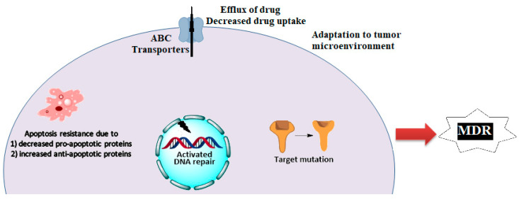
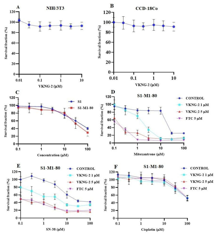
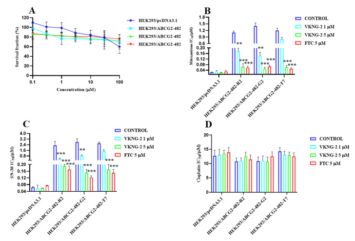
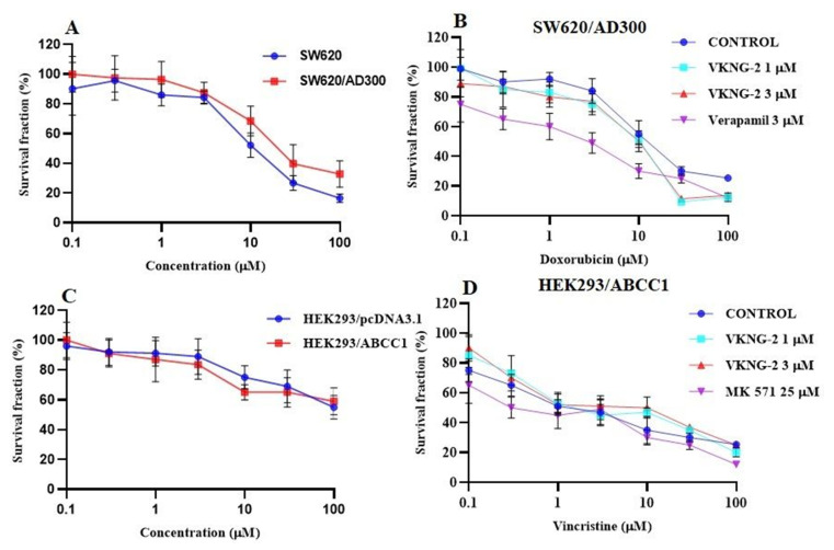
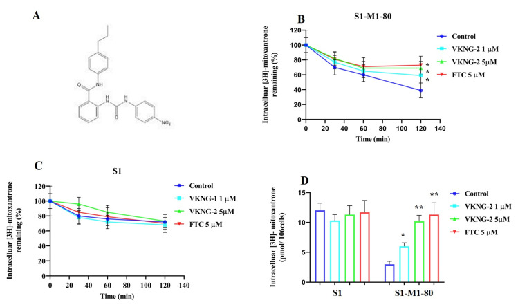
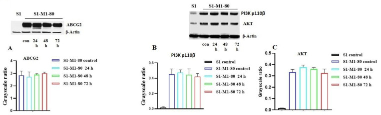
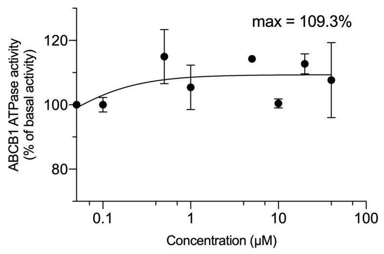
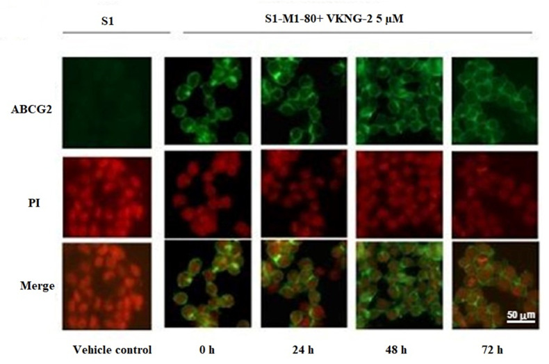
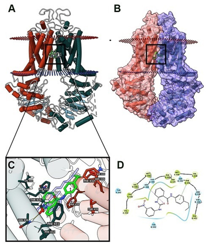
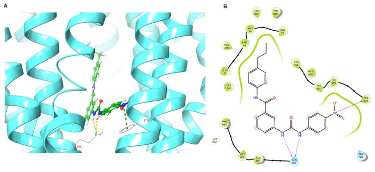
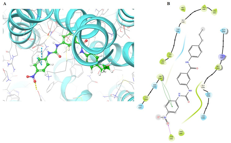
The overexpression of ATP-binding cassette transporter, ABCG2, plays an important role in mediating multidrug resistance (MDR) in certain types of cancer cells. ABCG2-mediated MDR can significantly attenuate or abrogate the efficacy of anticancer drugs by increasing their efflux from cancer cells. In this study, we determined the efficacy of the novel benzamide derivative, VKNG-2, to overcome MDR due to the overexpression of the ABCG2 transporter in the colon cancer cell line, S1-M1-80. In vitro, 5 μM of VKNG-2 reversed the resistance of S1-M1-80 cell line to mitoxantrone (70-fold increase in efficacy) or SN-38 (112-fold increase in efficacy). In contrast, in vitro, 5 μM of VKNG-2 did not significantly alter either the expression of ABCG2, AKT, and PI3K p110β protein or the subcellular localization of the ABCG2 protein compared to colon cancer cells incubated with the vehicle. Molecular docking data indicated that VKNG-2 had a high docking score (-10.2 kcal/mol) for the ABCG2 transporter substrate-drug binding site whereas it had a low affinity on ABCB1 and ABCC1 transporters. Finally, VKNG-2 produced a significant concentration-dependent increase in ATPase activity (EC50 = 2.3 µM). In conclusion, our study suggests that in vitro, VKNG-2 reverses the resistance of S1-M1-80, a cancer cell line resistant to mitoxantrone and SN-38, by inhibiting the efflux function of the ABCG2 transporter.

Keywords: ABCG2; ATP binding cassette; MDR; benzamide; reversal effect.


Conflict of interest statement

The authors declare no conflict of interest.

Figures

Scheme 1
Scheme 1
Mechanisms of Multidrug resistance in cancer.
Figure 1
Figure 1
The effect of VKNG-2 in transformed mouse fibroblast (NIH/3T3) and human colon fibroblast (CCD-18Co) cell lines (A,B). The effect of VKNG-2 in S1 parental and ABCG2-overexpressing S1-M1-80 colon cancer cells (C). The survival fraction (%) was determined following incubation with 5 µM of VKNG-2 for 72 h in S1 (blue) and S1-M1-80 (red) cell lines and IC50 values of mitoxantrone (D), SN-38 (E), and cisplatin (F) in parental S1 and drug-selected ABCG2 overexpressing S1-M1-80 colon cancer cells with or without VKNG-2. The points with error bars represent the mean ± SD of independent determinations in triplicate. The figures are representative of three independent experiments.
Figure 2
Figure 2
The effect of VKNG-2 in HEK293 cells transfected the gene coding for the ABCG2 transporter. (A) The survival fraction (%) for HEK293/pcDNA3.1 (empty DNA vector control), HEK293/ABCG2-482-R2, HEK293/ABCG2-482-G2, and HEK293/ABCG2-482-T7 cell lines was determined following incubation with 5 µM of VKNG-2 for 72 h. The IC50 values for mitoxantrone following incubation with VKNG-2 (1 or 5 µM) or FTC (5 µM) for 72 h in HEK293/pcDNA3.1 (empty DNA vector control), HEK293/ABCG2-482-R2 (HEK293/ABCG2-482-G2 and HEK293/ABCG2-482-T7 cell lines. The IC50 values of mitoxantrone (B), SN-38 (C), and cisplatin (D) in HEK293/pcDNA3.1 (empty DNA vector control), HEK293/ABCG2-482-R2, HEK293/ABCG2-482-G2, and HEK293/ABCG2-482-T7 cell lines. The points with error bars represent the mean ± SD of independent determinations in triplicate. The figures are representative of three independent experiments. ** p ≤ 0.01 and *** p < 0.001 compared to the control group.
Figure 3
Figure 3
The effect of VKNG-2 in SW620 parental, ABCB1-overexpressing SW620/Ad300 colon cancer cells and HEK293/pcDNA3.1 parental and HEK293/ABCC1 transfected cells. (A) The survival fraction (%) for the SW620 parental and SW620/Ad300 colon cancer cell lines were determined following incubation with VKNG-2 for 72 h. (B) The IC50 values of doxorubicin in the presence of vehicle (Control), VKNG-2 (1 or 3 μM) or verapamil (3 μM) for 72 h in SW620 parental and SW620/Ad300 colon cancer cells. (C): The survival fraction (%) for the HEK293/pcDNA3.1 (empty DNA vector control) and HEK293/ABCC1 (transfected with the DNA coding for the ABCC1 transporter) cells were determined following incubation with VKNG-2 for 72 h. (D) The IC50 values of vincristine in the presence of vehicle (Control), VKNG-2 (1 or 3 μM) or MK-571 (25 μM) for 72 h in HEK293/pcDNA3.1 and HEK293/ABCC1 cells. The points with error bars represent the mean ± SD of independent determinations in triplicate. The figures are representative of three independent experiments.
Figure 4
Figure 4
(A) The chemical structure of VKNG-2. (B) The effect of the incubation of vehicle (Control), VKNG-2 (1 or 5 µM) or FTC (5 µM) for 30, 60 or 120 min on the efflux of the ABCG2 transporter substrate, [3H]-mitoxantrone from S1-M1-80 colon cancer cells overexpressing the ABCG2 transporter. (C) The effect of the incubation of vehicle (Control), VKNG-2 (1 or 5 µM) or FTC (5 µM) for 30, 60 or 120 min on the efflux of the ABCG2 transporter substrate, [3H]-mitoxantrone from S1 parental colon cancer cells. (D) The effect of the vehicle (Control), VKNG-2 (1 or 5 µM) or FTC (5 µM) on the intracellular accumulation of [3H]-mitoxantrone in S1 and S1-M1-80 colon cancer cells. The columns are the mean of triplicate determinations; the error bars represent the SD. * p ≤ 0.05 and ** p ≤ 0.01 compared with the control group.
Figure 5
Figure 5
The effect of VKNG-2 on the expression of the ABCG2, PI3K p110β and AKT protein. The effect of VKNG-2 on the expression of the ABCG2, PI3K p110β and AKT protein were determined in S1-M1-80 colon cancer cells following incubation with vehicle (Control) or 5 μM of VKNG-2 for 24, 48 or 72 h (AC). Equal amounts of total cell lysates were used for each sample and a Western blot analysis was performed.
Figure 6
Figure 6
VKNG-2 stimulated the ATPase activity of ABCB1. The graph illustrates the effect of 0–100 µM of VKNG-2 on the ATPase activity of ABCB1.
Figure 7
Figure 7
The effect of VKNG-2 on the expression and localization of ABCG2 using immunofluorescence. The effect of the incubation of S1 and S1-M1-80 colon cancer cells with vehicle (Control) or 5 µM of VKNG-2 for incubated for 0, 24, 48 or 72 h. The green color represents the presence of the ABCG2 transporter, and the red color represents the nucleus.
Figure 8
Figure 8
The molecular modeling of VKNG-2 and human ABCG2. (A) An overview of mitoxantrone and the best-scoring pose of VKNG-2 in the drug-substrate binding site of the ABCG2 protein (6VXI). The cytoplasm membrane is depicted as dotted planes, where the red or blue planes indicate the extracellular or intracellular side, respectively. ABCG2 is displayed as colored tubes and ribbons. VKNG-2 and mitoxantrone are displayed as colored sticks. Carbon: lime green (VKNG-2) or white (mitoxantrone); oxygen: red; nitrogen: blue. (B) Mitoxantrone and the best-scoring pose of VKNG-2 in the drug-substrate binding site of the ABCG2 protein with molecule surface displayed. (C) Details of the interactions between VKNG-2 and the ABCG2 (6VXI) drug-substrate binding site. ABCG2 protein helices are displayed as colored tubes (chain A: green; chain B: red). Important residues are displayed as colored sticks (carbon: same as chain color; oxygen: red; nitrogen: blue). VKNG-2 is displayed as colored sticks (carbon: lime; oxygen: red; nitrogen: blue). Hydrogen bonds are displayed as yellow dash lines. p-p stacking interactions are displayed as magenta dash lines. (D) 2D diagram of the interaction between VKNG-2 and ABCG2. Important amino acids are displayed as colored bubbles (green: hydrophobic; blue: polar). Purple solid lines with an arrow indicate hydrogen bonds. Green solid lines without arrow indicate p-p stacking interactions.
Figure 9
Figure 9
Molecular interaction of VKNG-2 with the human ABCB1 model. (A) Docking pose of VKNG-2 within the binding pocket of ABCB1. The protein is represented as sky blue colored ribbons. Amino acid residues are represented as follows: carbon in gray, hydrogen in white, nitrogen in blue and oxygen in red. The ligand is represented by the ball and stick model with carbon atoms represented in green, oxygen in red nitrogen in blue and hydrogen in white. Green dashes represent π-cation interactions and yellow dashes represent hydrogen bonding. (B) 2-D ligand interaction between VKNG-2 and ABCB1. Red arrows indicate π-cation interaction with amino acid residues within 5 Å of the ligand and the magenta arrows represent hydrogen bonding.
Figure 10
Figure 10
Molecular interaction of VKNG-2 with the human ABCC1 model. (A) Docking pose of VKNG-2 within the binding pocket of ABCC1. The protein is represented as sky blue colored ribbons. Amino acid residues are represented as follows: carbon in gray, hydrogen in white, nitrogen in blue and oxygen in red. The ligand is represented by the ball and stick model with carbon atoms represented in green, oxygen in red nitrogen in blue and hydrogen in white. Blue dashes represent π-π stacking interaction, yellow dashes represent the hydrogen bonding. (B) 2-D ligand interaction between VKNG-2 and ABCC1. Green indicates π-π interaction with amino acid residues within 5 Å of the ligand and the magenta arrow represents hydrogen bonding.

Similar articles

Cited by

References

    1. Narayanan S., Gupta P., Nazim U., Ali M., Karadkhelkar N., Ahmad M., Chen Z.-S. Anti-cancer effect of Indanone-based thiazolyl hydrazone derivative on colon cancer cell lines. Int. J. Biochem. Cell Biol. 2019;110:21–28. doi: 10.1016/j.biocel.2019.02.004. - DOI - PubMed
    1. Barbuti A.M., Zhang G.-N., Gupta P., Narayanan S., Chen Z.-S. Protein Kinase Inhibitors as Sensitizing Agents for Chemotherapy. Volume 4. Elsevier; Amsterdam, The Netherlands: 2019. EGFR and HER2 Inhibitors as Sensitizing Agents for Cancer Chemotherapy; pp. 1–11.
    1. Gupta P., Narayanan S., Yang D.-H. Protein Kinase Inhibitors as Sensitizing Agents for Chemotherapy. Volume 4. Elsevier; Amsterdam, The Netherlands: 2019. CDK Inhibitors as Sensitizing Agents for Cancer Chemotherapy; pp. 125–149.
    1. Bray F., Ferlay J., Soerjomataram I., Siegel R.L., Torre L.A., Jemal A. Global cancer statistics 2018: GLOBOCAN estimates of incidence and mortality worldwide for 36 cancers in 185 countries. CA Cancer J. Clin. 2018;68:394–424. doi: 10.3322/caac.21492. - DOI - PubMed
    1. Narayanan S., Cai C.-Y., Assaraf Y.G., Guo H.-Q., Cui Q., Wei L., Huang J.-J., Ashby C.R., Chen Z.-S. Targeting the ubiquitin-proteasome pathway to overcome anti-cancer drug resistance. Drug Resist. Updat. 2020;48:100663. doi: 10.1016/j.drup.2019.100663. - DOI - PubMed

MeSH terms

LinkOut - more resources