

Oxidative Stress and ROS-Mediated Signaling in Leukemia: Novel Promising Perspectives to Eradicate Chemoresistant Cells in Myeloid Leukemia
- PMID: 33671113
- PMCID: PMC7957553
- DOI: 10.3390/ijms22052470
Oxidative Stress and ROS-Mediated Signaling in Leukemia: Novel Promising Perspectives to Eradicate Chemoresistant Cells in Myeloid Leukemia
Abstract
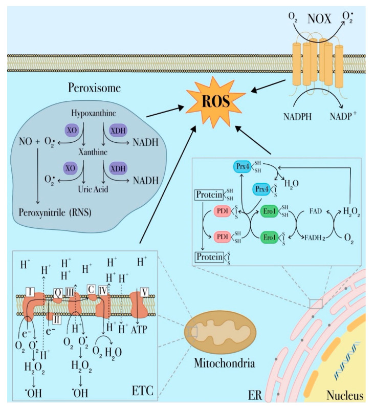
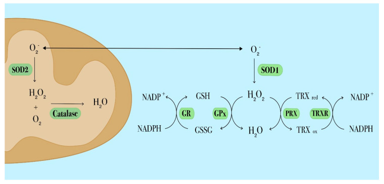
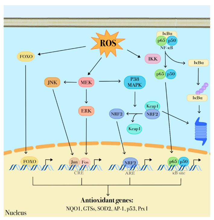
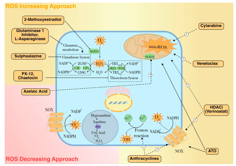
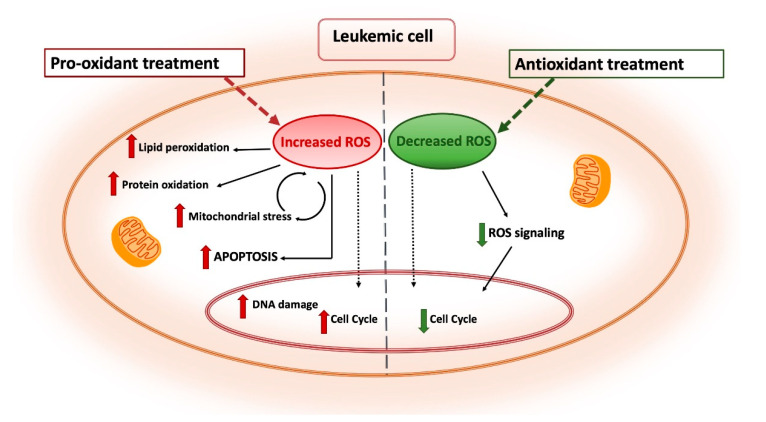
Myeloid leukemic cells are intrinsically under oxidative stress due to impaired reactive oxygen species (ROS) homeostasis, a common signature of several hematological malignancies. The present review focuses on the molecular mechanisms of aberrant ROS production in myeloid leukemia cells as well as on the redox-dependent signaling pathways involved in the leukemogenic process. Finally, the relevance of new chemotherapy options that specifically exert their pharmacological activity by altering the cellular redox imbalance will be discussed as an effective strategy to eradicate chemoresistant cells.
Keywords: ROS; ROS-based therapy; acute myeloid leukemia (AML); antioxidant systems; chemoreistance; oxidative stress.
Conflict of interest statement
The authors declare that they have no conflict of interest.
Figures
Similar articles
-
Targeting Myeloperoxidase Disrupts Mitochondrial Redox Balance and Overcomes Cytarabine Resistance in Human Acute Myeloid Leukemia.Cancer Res. 2019 Oct 15;79(20):5191-5203. doi: 10.1158/0008-5472.CAN-19-0515. Epub 2019 Jul 29. Cancer Res. 2019. PMID: 31358527
-
Oxidative stress is two-sided in the treatment of acute myeloid leukemia.Cancer Med. 2024 May;13(9):e6806. doi: 10.1002/cam4.6806. Cancer Med. 2024. PMID: 38715546 Free PMC article. Review.
-
Peperomin E and its orally bioavailable analog induce oxidative stress-mediated apoptosis of acute myeloid leukemia progenitor cells by targeting thioredoxin reductase.Redox Biol. 2019 Jun;24:101153. doi: 10.1016/j.redox.2019.101153. Epub 2019 Mar 8. Redox Biol. 2019. PMID: 30909158 Free PMC article.
-
Reactive oxygen species activate differentiation gene transcription of acute myeloid leukemia cells via the JNK/c-JUN signaling pathway.Leuk Res. 2018 May;68:112-119. doi: 10.1016/j.leukres.2018.03.012. Epub 2018 Mar 27. Leuk Res. 2018. PMID: 29609096
-
Harnessing Azelaic Acid for Acute Myeloid Leukemia Treatment: A Novel Approach to Overcoming Chemoresistance and Improving Outcomes.Int J Mol Sci. 2025 May 3;26(9):4362. doi: 10.3390/ijms26094362. Int J Mol Sci. 2025. PMID: 40362596 Free PMC article. Review.
Cited by
-
A multidisciplinary approach disclosing unexplored Aflatoxin B1 roles in severe impairment of vitamin D mechanisms of action.Cell Biol Toxicol. 2023 Aug;39(4):1275-1295. doi: 10.1007/s10565-022-09752-y. Epub 2022 Sep 6. Cell Biol Toxicol. 2023. PMID: 36066700 Free PMC article.
-
Oral issues and childhood stress in eight-to-ten-year-old schoolchildren: a case-control study.Clin Oral Investig. 2024 Aug 31;28(9):509. doi: 10.1007/s00784-024-05889-8. Clin Oral Investig. 2024. PMID: 39215814
-
Emerging role of glutathione peroxidase 4 in myeloid cell lineage development and acute myeloid leukemia.Cell Mol Biol Lett. 2024 Jul 8;29(1):98. doi: 10.1186/s11658-024-00613-6. Cell Mol Biol Lett. 2024. PMID: 38977956 Free PMC article. Review.
-
Signaling pathways governing the behaviors of leukemia stem cells.Genes Dis. 2023 Mar 23;11(2):830-846. doi: 10.1016/j.gendis.2023.01.008. eCollection 2024 Mar. Genes Dis. 2023. PMID: 37692500 Free PMC article. Review.
-
Over-Expressed GATA-1S, the Short Isoform of the Hematopoietic Transcriptional Factor GATA-1, Inhibits Ferroptosis in K562 Myeloid Leukemia Cells by Preventing Lipid Peroxidation.Antioxidants (Basel). 2023 Feb 21;12(3):537. doi: 10.3390/antiox12030537. Antioxidants (Basel). 2023. PMID: 36978786 Free PMC article.
References
-
- Sies H. Stress: Physiology, Biochemistry, and Pathology. Elsevier; Amsterdam, The Netherlands: 2019. Oxidative Stress: Eustress and Distress in Redox Homeostasis; pp. 153–163.
Publication types
MeSH terms
Substances
Grants and funding
LinkOut - more resources
Full Text Sources
Other Literature Sources
Medical
