

How miR-31-5p and miR-33a-5p Regulates SP1/CX43 Expression in Osteoarthritis Disease: Preliminary Insights
- PMID: 33671114
- PMCID: PMC7957523
- DOI: 10.3390/ijms22052471
How miR-31-5p and miR-33a-5p Regulates SP1/CX43 Expression in Osteoarthritis Disease: Preliminary Insights
Abstract
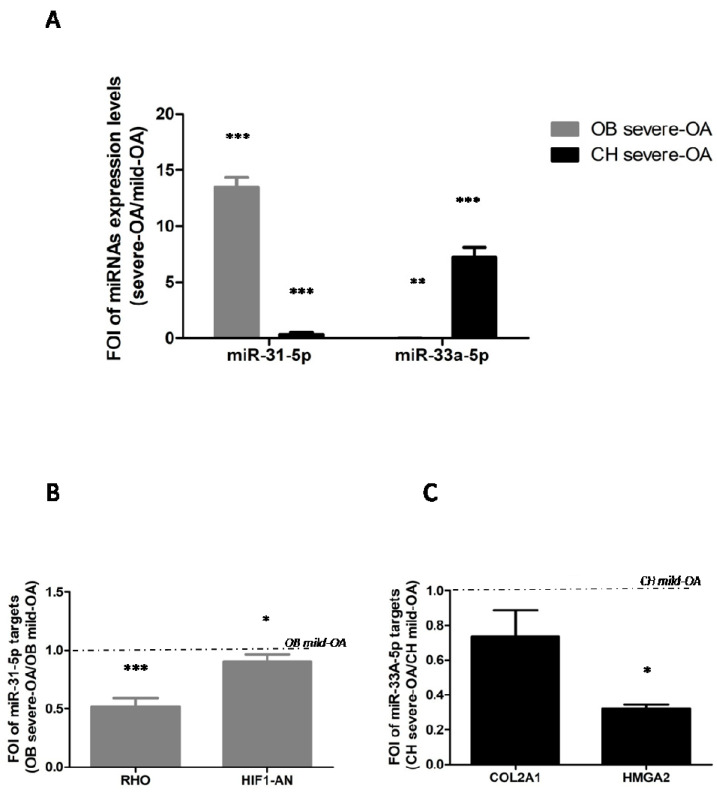
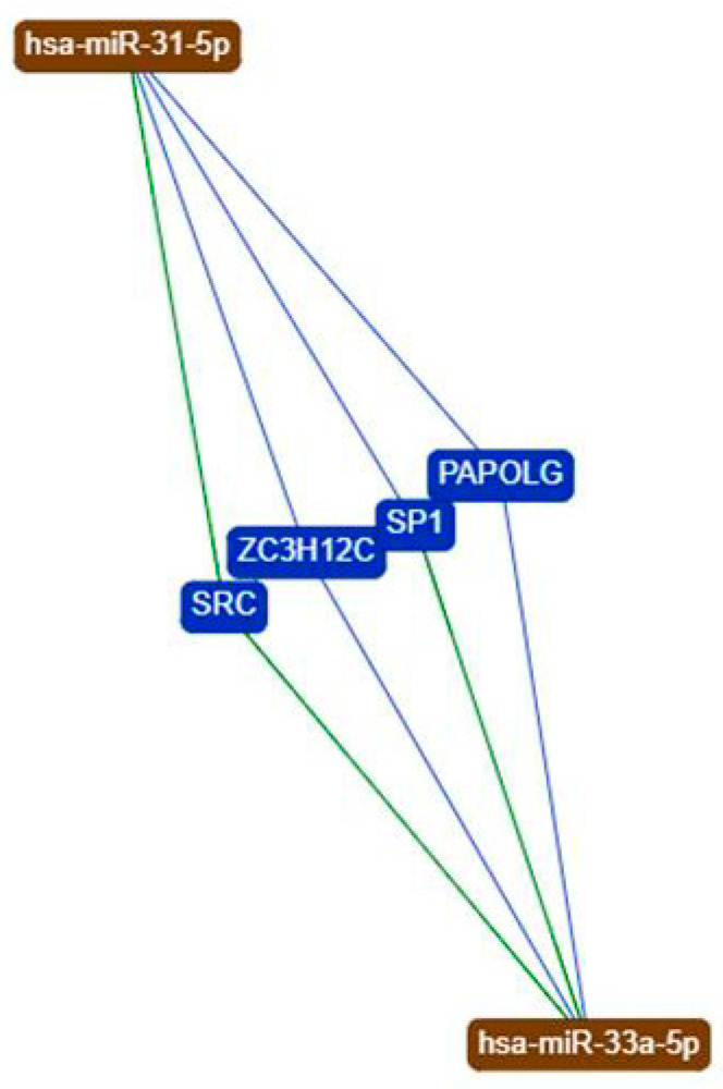
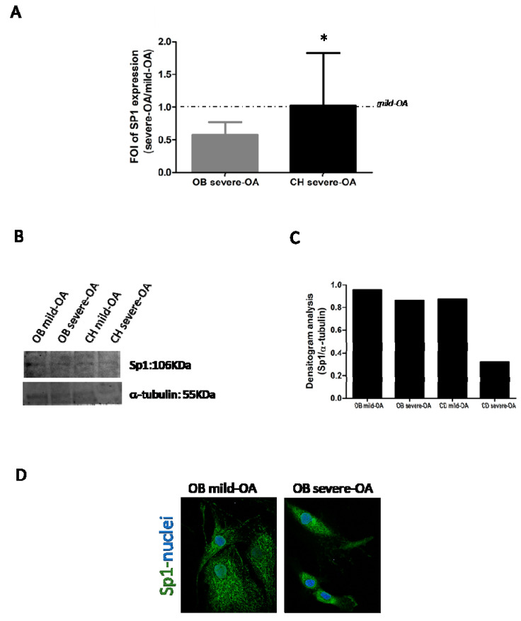
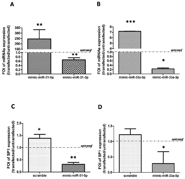
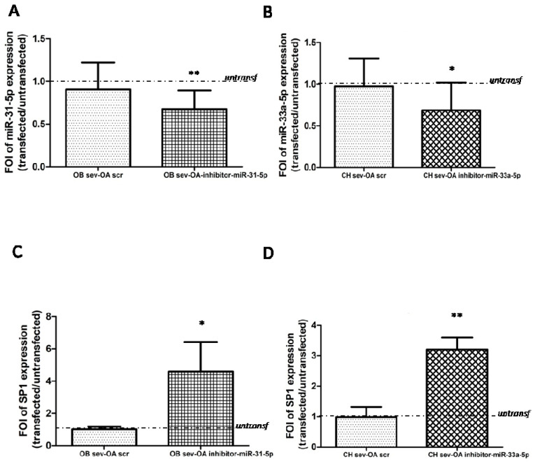
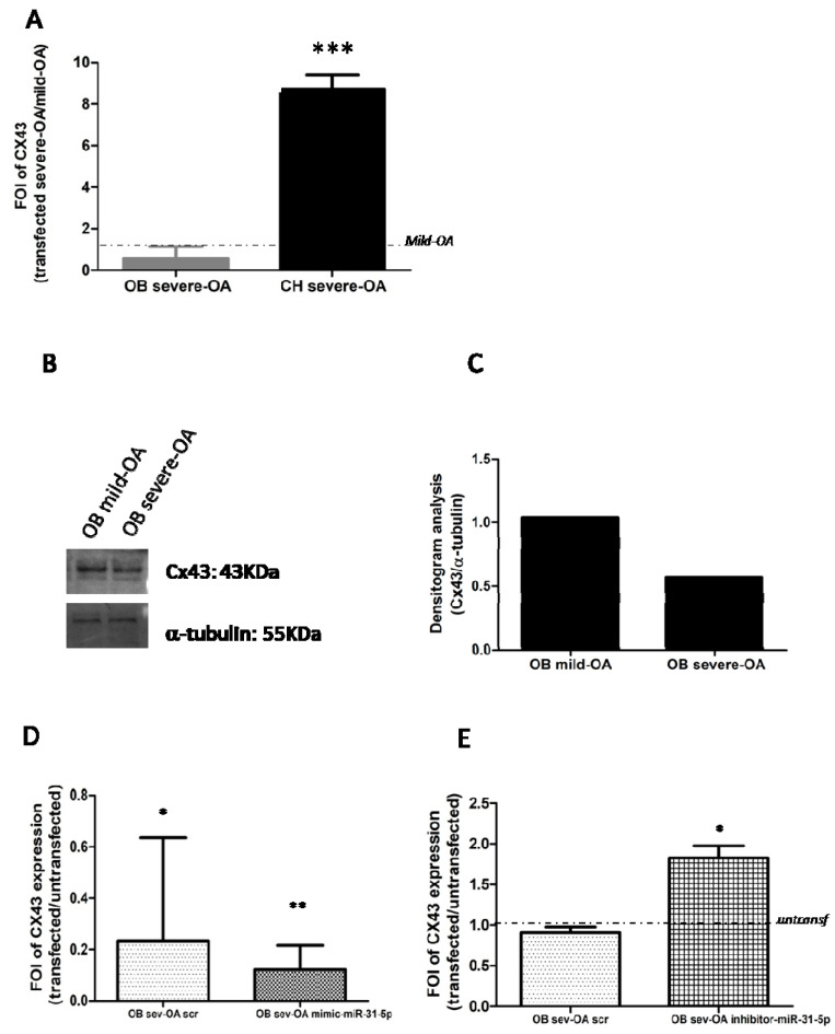
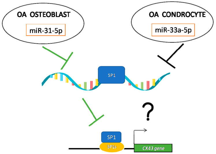
Osteoarthritis (OA) is a degenerative bone disease that involved micro and macro-environment of joints. To date, there are no radical curative treatments for OA and novel therapies are mandatory. Recent evidence suggests the role of miRNAs in OA progression. In our previous studies, we demonstrated the role of miR-31-5p and miR-33a families in different bone regeneration signaling. Here, we investigated the role of miR-31-5p and miR-33a-5p in OA progression. A different expression of miR-31-5p and miR-33a-5p into osteoblasts and chondrocytes isolated from joint tissues of OA patients classified in based on different Kellgren and Lawrence (KL) grading was highlighted; and through a bioinformatic approach the common miRNAs target Specificity proteins (Sp1) were identified. Sp1 regulates the expression of gap junction protein Connexin43 (Cx43), which in OA drives the modification of i) osteoblasts and chondrocytes genes expression, ii) joint inflammation cytokines releases and iii) cell functions. Concerning this, thanks to gain and loss of function studies, the possible role of Sp1 as a modulator of CX43 expression through miR-31-5p and miR-33a-5p action was also evaluated. Finally, we hypothesize that both miRNAs cooperate to modulate the expression of SP1 in osteoblasts and chondrocytes and interfering, consequently, with CX43 expression, and they might be further investigated as new possible biomarkers for OA.
Keywords: CX43; SP1; chondrocytes; microRNAs; osteoarthritis; osteoblasts.
Conflict of interest statement
The authors declare no conflict of interest.
Figures
Similar articles
-
DNA methylation regulates miR-140-5p and miR-146a expression in osteoarthritis.Life Sci. 2019 Jul 1;228:274-284. doi: 10.1016/j.lfs.2019.05.018. Epub 2019 May 9. Life Sci. 2019. PMID: 31077718
-
Positive Feedback Loop LINC00511/miR-150-5p/SP1 Modulates Chondrocyte Apoptosis and Proliferation in Osteoarthritis.DNA Cell Biol. 2020 Sep;39(9):1506-1512. doi: 10.1089/dna.2020.5718. Epub 2020 Jul 7. DNA Cell Biol. 2020. PMID: 32635763
-
Long non-coding RNA DANCR regulates proliferation and apoptosis of chondrocytes in osteoarthritis via miR-216a-5p-JAK2-STAT3 axis.Biosci Rep. 2018 Dec 14;38(6):BSR20181228. doi: 10.1042/BSR20181228. Print 2018 Dec 21. Biosci Rep. 2018. PMID: 30361290 Free PMC article.
-
MicroRNA expression in osteoarthritis: a meta-analysis.Clin Exp Med. 2023 Nov;23(7):3737-3749. doi: 10.1007/s10238-023-01063-8. Epub 2023 Apr 7. Clin Exp Med. 2023. PMID: 37027064
-
Relationship between Oxytocin and Osteoarthritis: Hope or Despair?Int J Mol Sci. 2021 Oct 29;22(21):11784. doi: 10.3390/ijms222111784. Int J Mol Sci. 2021. PMID: 34769215 Free PMC article. Review.
Cited by
-
Mechanism of miRNA-31 Regulating Wnt/β-catenin Signaling Pathway by Targeting Satb2 in the Osteogenic Differentiation of Human Bone Marrow-Derived Mesenchymal Stem Cells.J Musculoskelet Neuronal Interact. 2023 Sep 1;23(3):346-354. J Musculoskelet Neuronal Interact. 2023. PMID: 37654220 Free PMC article.
-
Profiling Blood Serum Extracellular Vesicles in Plaque Psoriasis and Psoriatic Arthritis Patients Reveals Potential Disease Biomarkers.Int J Mol Sci. 2022 Apr 4;23(7):4005. doi: 10.3390/ijms23074005. Int J Mol Sci. 2022. PMID: 35409365 Free PMC article.
-
MiR203a-3p as a potential biomarker for synovial pathology associated with osteoarthritis: a pilot study.J Orthop Surg Res. 2024 Nov 12;19(1):746. doi: 10.1186/s13018-024-05237-2. J Orthop Surg Res. 2024. PMID: 39533317 Free PMC article.
-
Adipose tissue-derived versus bone marrow-derived cell concentrates for the injective treatment of knee osteoarthritis: protocol of a prospective randomised controlled trial.BMJ Open. 2025 Apr 30;15(4):e092379. doi: 10.1136/bmjopen-2024-092379. BMJ Open. 2025. PMID: 40306987 Free PMC article.
-
Perspective and Therapeutic Potential of the Noncoding RNA-Connexin Axis.Int J Mol Sci. 2024 Jun 2;25(11):6146. doi: 10.3390/ijms25116146. Int J Mol Sci. 2024. PMID: 38892334 Free PMC article. Review.
References
-
- Gibon E., Córdova L.A., Lu L., Lin T.-H., Yao Z., Hamadouche M., Goodman S.B. The biological response to orthopedic implants for joint replacement. II: Polyethylene, ceramics, PMMA, and the foreign body reaction. J. Biomed. Mater. Res. Part B Appl. Biomater. 2017;105:1685–1691. doi: 10.1002/jbm.b.33676. - DOI - PMC - PubMed
MeSH terms
Substances
LinkOut - more resources
Full Text Sources
Other Literature Sources
Medical
Miscellaneous
