

Mycobacterium tuberculosis Small RNA MTS1338 Confers Pathogenic Properties to Non-Pathogenic Mycobacterium smegmatis
- PMID: 33671144
- PMCID: PMC7921967
- DOI: 10.3390/microorganisms9020414
Mycobacterium tuberculosis Small RNA MTS1338 Confers Pathogenic Properties to Non-Pathogenic Mycobacterium smegmatis
Abstract
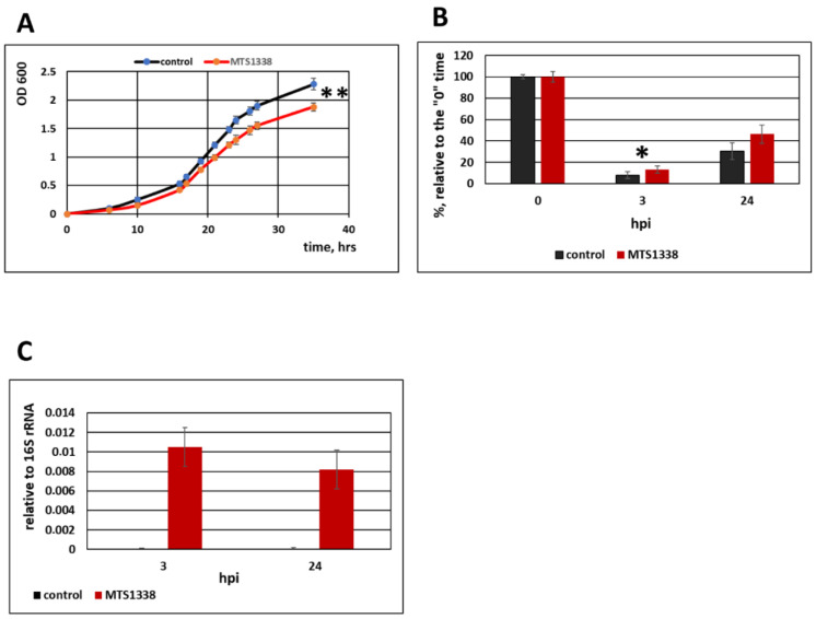
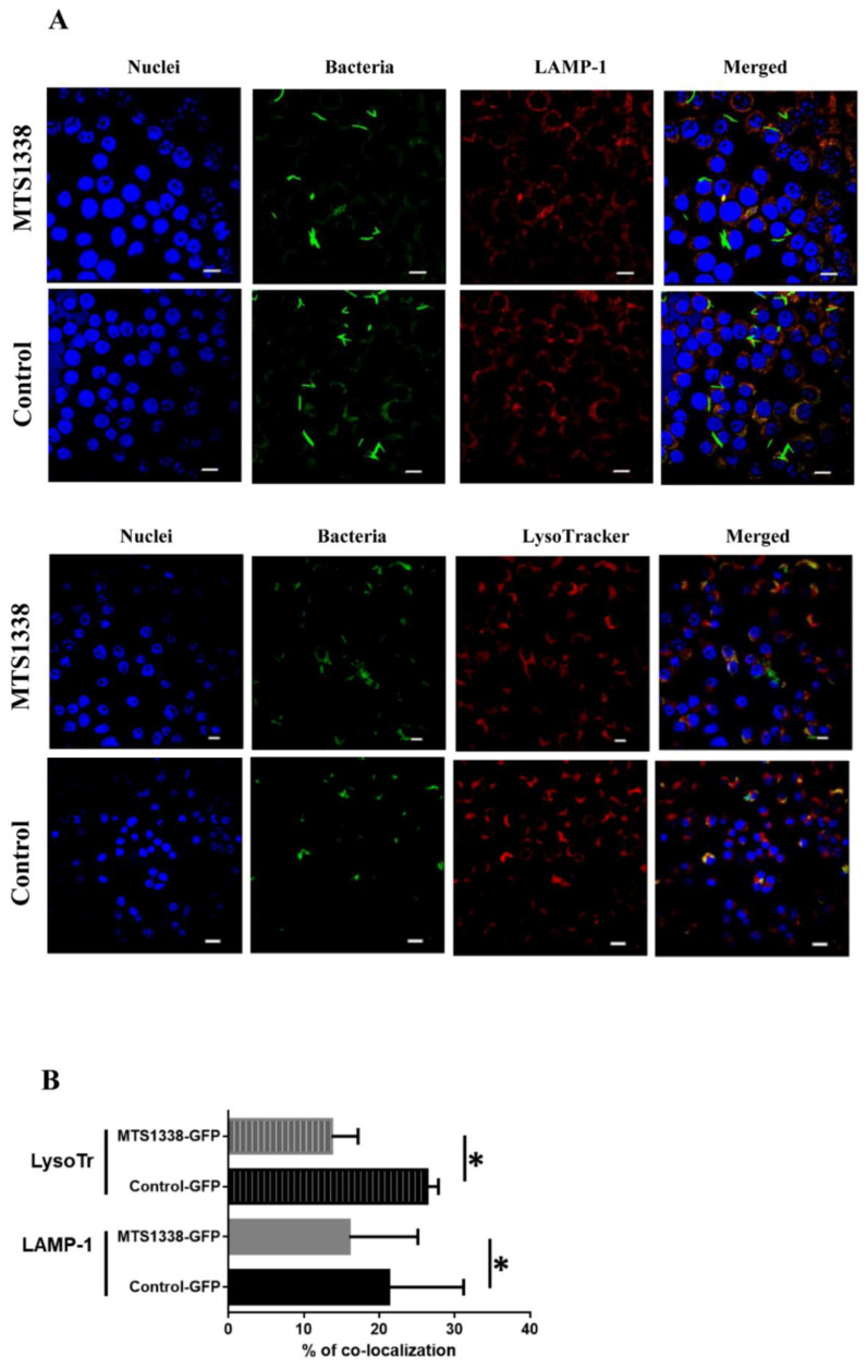
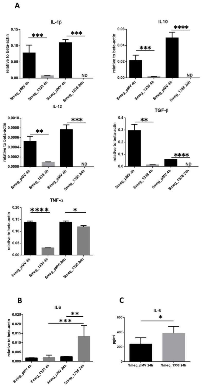
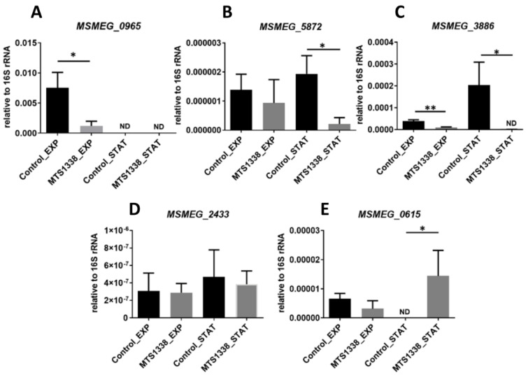
Small non-coding RNAs play a key role in bacterial adaptation to various stresses. Mycobacterium tuberculosis small RNA MTS1338 is upregulated during mycobacteria infection of macrophages, suggesting its involvement in the interaction of the pathogen with the host. In this study, we explored the functional effects of MTS1338 by expressing it in non-pathogenic Mycobacterium smegmatis that lacks the MTS1338 gene. The results indicated that MTS1338 slowed the growth of the recombinant mycobacteria in culture and increased their survival in RAW 264.7 macrophages, where the MTS1338-expressing strain significantly (p < 0.05) reduced the number of mature phagolysosomes and changed the production of cytokines IL-1β, IL-6, IL-10, IL-12, TGF-β, and TNF-α compared to those of the control strain. Proteomic and secretomic profiling of recombinant and control strains revealed differential expression of proteins involved in the synthesis of main cell wall components and in the regulation of iron metabolism (ESX-3 secretion system) and response to hypoxia (furA, whiB4, phoP). These effects of MTS1338 expression are characteristic for M. tuberculosis during infection, suggesting that in pathogenic mycobacteria MTS1338 plays the role of a virulence factor supporting the residence of M. tuberculosis in the host.
Keywords: MTS1338; Mycobacterium smegmatis; Mycobacterium tuberculosis; macrophages; proteomics; small non-coding RNA; virulence factor.
Conflict of interest statement
The authors declare no conflict of interest.
Figures
Similar articles
-
Small RNA MTS1338 Configures a Stress Resistance Signature in Mycobacterium tuberculosis.Int J Mol Sci. 2023 Apr 27;24(9):7928. doi: 10.3390/ijms24097928. Int J Mol Sci. 2023. PMID: 37175635 Free PMC article.
-
MTS1338, A Small Mycobacterium tuberculosis RNA, Regulates Transcriptional Shifts Consistent With Bacterial Adaptation for Entering Into Dormancy and Survival Within Host Macrophages.Front Cell Infect Microbiol. 2019 Nov 26;9:405. doi: 10.3389/fcimb.2019.00405. eCollection 2019. Front Cell Infect Microbiol. 2019. PMID: 31850238 Free PMC article.
-
Transcriptional activation of the Mycobacterium tuberculosis virulence-associated small RNA MTS1338 by the response regulators DosR and PhoP.FEBS Lett. 2024 May;598(9):1034-1044. doi: 10.1002/1873-3468.14882. Epub 2024 Apr 19. FEBS Lett. 2024. PMID: 38639734
-
[Systems of genes and proteins affecting mycobacteria virulence and their homologs participation in conjugation of mycobacterium smegmatis].Genetika. 2013 Jan;49(1):125-41. doi: 10.7868/s0016675813010098. Genetika. 2013. PMID: 23662430 Review. Russian.
-
Comparing the Metabolic Capabilities of Bacteria in the Mycobacterium tuberculosis Complex.Microorganisms. 2019 Jun 18;7(6):177. doi: 10.3390/microorganisms7060177. Microorganisms. 2019. PMID: 31216777 Free PMC article. Review.
Cited by
-
Red light-emitting short Mango-based system enables tracking a mycobacterial small noncoding RNA in infected macrophages.Nucleic Acids Res. 2023 Apr 11;51(6):2586-2601. doi: 10.1093/nar/gkad100. Nucleic Acids Res. 2023. PMID: 36840712 Free PMC article.
-
Pyridine Compounds with Antimicrobial and Antiviral Activities.Int J Mol Sci. 2022 May 18;23(10):5659. doi: 10.3390/ijms23105659. Int J Mol Sci. 2022. PMID: 35628466 Free PMC article. Review.
-
Small RNA MTS1338 Configures a Stress Resistance Signature in Mycobacterium tuberculosis.Int J Mol Sci. 2023 Apr 27;24(9):7928. doi: 10.3390/ijms24097928. Int J Mol Sci. 2023. PMID: 37175635 Free PMC article.
-
Small RNA Profiling in Mycobacterium Provides Insights Into Stress Adaptability.Front Microbiol. 2021 Nov 4;12:752537. doi: 10.3389/fmicb.2021.752537. eCollection 2021. Front Microbiol. 2021. PMID: 34803973 Free PMC article.
-
A virulence-associated small RNA MTS1338 activates an ABC transporter CydC for rifampicin efflux in Mycobacterium tuberculosis.Front Microbiol. 2024 Sep 19;15:1469280. doi: 10.3389/fmicb.2024.1469280. eCollection 2024. Front Microbiol. 2024. PMID: 39364170 Free PMC article.
References
-
- Deretic V., Singh S., Master S., Harris J., Roberts E., Kyei G., Davis A., de Haro S., Naylor J., Lee H.H., et al. Mycobacterium tuberculosis inhibition of phagolysosome biogenesis and autophagy as a host defence mechanism. Cell Microbiol. 2006;8:719–727. doi: 10.1111/j.1462-5822.2006.00705.x. - DOI - PubMed
Grants and funding
LinkOut - more resources
Full Text Sources
Other Literature Sources
