. 2021 Feb 17;10(2):307.
doi: 10.3390/antiox10020307.

Inhibitory Effect of a Rosmarinic Acid-Enriched Fraction Prepared from Nga-Mon (Perilla frutescens) Seed Meal on Osteoclastogenesis through the RANK Signaling Pathway

Affiliations

Inhibitory Effect of a Rosmarinic Acid-Enriched Fraction Prepared from Nga-Mon (Perilla frutescens) Seed Meal on Osteoclastogenesis through the RANK Signaling Pathway

Kanokkarn Phromnoi et al. Antioxidants (Basel). .

Abstract

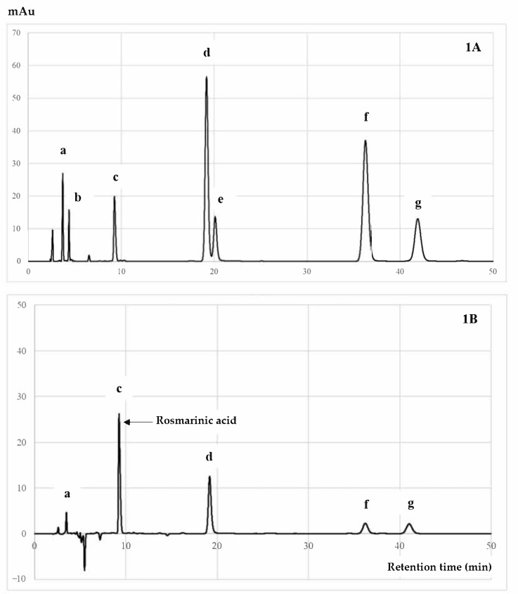
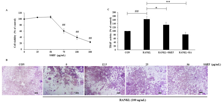
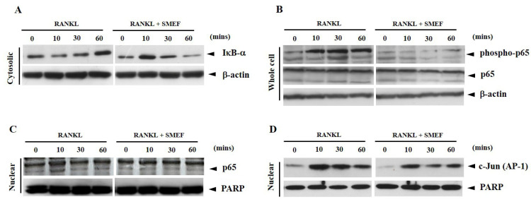
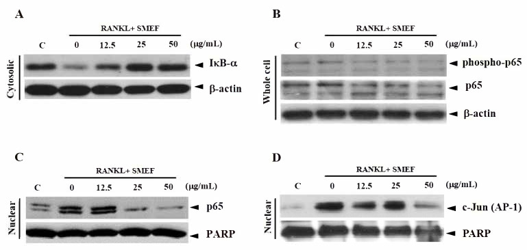
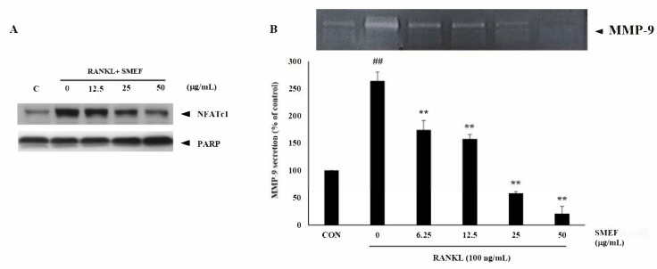
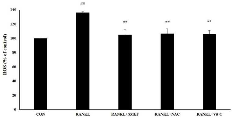
The aim of this study is to determine antioxidant and anti-inflammatory activities relating to the antiosteoporosis effects of various perilla seed meal (PSM) fractions. The remaining waste of perilla seed obtained from cold oil compression was extracted with 70% ethanol and sequentially fractionated according to solvent polarity with hexane, dichloromethane, ethyl acetate, and water. The results indicated that the seed-meal ethyl acetate fraction (SMEF) exhibited the highest antioxidant and anti-inflammatory activities, and rosmarinic acid (RA) content. The signaling pathways induced by the receptor activator of the nuclear factor kappa B (NF-κB) ligand (RANKL) that trigger reactive oxygen species (ROS) and several transcription factors, leading to the induction of osteoclastogenesis, were also investigated. The SMEF clearly showed attenuated RANKL-induced tartrate-resistant acid phosphatase (TRAP)-positive multinucleated osteoclasts and TRAP activity. A Western blot analysis showed that the SMEF significantly downregulated RANKL-induced NF-κB, AP-1 activation, and the nuclear factor of activated T-cell 1 (NFATc1) expression. SMEF also suppressed RANKL-induced osteoclast-specific marker gene-like MMP-9 using zymography. Furthermore, the SMEF showed inhibition of RANKL-induced ROS production in RAW 264.7 cells. The results suggest that the SMEF, which contained high quantities of RA, could be developed as a natural active pharmaceutical ingredient for osteoclastogenic protection and health promotion.

Keywords: Nga-Mon (Perilla frutescens); RANKL; ROS; anti-inflammation; antioxidant; osteoclastogenesis.


Conflict of interest statement

The authors declare no conflict of interest.

Figures

Figure 1
Figure 1
HPLC chromatogram of (A) mixed standards and (B) SMEF: (a) caffeic acid; (b) rutin; (c) rosmarinic acid; (d) luteolin; (e) quercetin; (f) apigenin; (g) kaempferol.
Figure 2
Figure 2
SMEF suppresses osteoclastogenesis in vitro. (A) RAW264.7 cells cultured with SMEF in concentration range of 0–200 μg/mL for 48 h in 96-well plates. Cell viability was determined by an MTT assay. ## p < 0.001 compared with untreated control. (B) RAW264.7 cells were coincubated with SMEF (0–50 μg/mL) and RANKL (100 ng/mL) for 6 days and then stained using a leukocyte acid phosphatase (TRAP) kit. TRAP-positive multinucleated osteoclasts were visualized in 100× magnification under light microphotography. Scale bars, 100.2 μm. (C) RAW264.7 cells were co-incubated with SMEF (50 μg/mL) and RANKL (100 ng/mL) for 6 day and then measured using the TRAP solution assay. RA (100 μM) was used as a positive control and TRAP activity was expressed as % of control. The data is presented as the mean ± SD of three independent experiments. ## p < 0.001, compared with control (CON); * p < 0.01, ** p < 0.001 compared with RANKL control.
Figure 3
Figure 3
SMEF suppresses RANKL-induced NF-κB and AP-1 activation after inducing with RANKL for indicated time. (AD) RAW 264.7 cells were pretreated with SMEF (50 μg/mL) for 12 h and then exposed to RANKL for 0, 10, 30, and 60 min. Cytosolic fractions were analyzed for protein content of IκB-α degradation, whole-cell lysates were used to determine phosphorylation levels of NF-κB p65, and nuclear fractions were analyzed for NF-κB p65 and AP-1(c-Jun) translocation (assayed by Western blotting as described in the Materials and Methods section).
Figure 4
Figure 4
SMEF suppresses RANKL-induced NF-κB and AP-1 activation in dose-dependent manner. (AD) RAW 264.7 cells were pretreated with SMEF (0, 12.5, 25, and 50 µg/mL) for 12 h and then exposed to RANKL for 10 min. Cytosolic fractions were analyzed for protein content of IκB-α degradation, whole-cell lysates were used to determine phosphorylation levels of NF-κB p65, and nuclear fractions were analyzed for NF-κB p65 and AP-1(c-Jun) translocation (assayed by Western blotting as described in the Materials and Methods section).
Figure 5
Figure 5
SMEF inhibits osteoclastic-specific proteins, NFATc1 and MMP-9 expression. (A) RAW264.7 cells were pretreated with SMEF in concentrations of 0, 12.5, 25, and 50 μg/mL for 12 h and stimulated by RANKL (100 ng/mL) for 10 min. Nuclear extracts were prepared and analyzed by Western blot analysis. (B) RAW264.7 cells were cotreated with SMEF in concentrations of 0, 12.5, 25, and 50 μg/mL and RANKL (100 ng/mL) for 2 days. Culture supernatants of treated cells were collected, and secretion of MMP-9 was analyzed by gelatin zymography. Data are presented as mean ± SD of three independent experiments. ## p < 0.001 compared with control (CON); ** p < 0.001 compared with RANKL control.
Figure 6
Figure 6
SMEF inhibits RANKL-induced reactive oxygen species (ROS) production using cellular dichlorodihydrofluorescein diacetate (DCF-DA) assay. RAW 264.7 cells were seeded in a 96-well plate, pretreated with the SMEF (50 µg/mL) for 24 h, and stimulated by RANKL (100 ng/mL) for 1 h. Then, 40 µM of DCFH-DA solution was added for 30 min. Treated cells were measured with a fluorescent microplate reader. Then, 50 µM N-acetylcysteine (NAC) and 250 µM ascorbic acid (Vit C) were used as a positive control. All data are presented as the mean ± SD of three independent experiments. ## p < 0.001 compared with the untreated control (CON); ** p < 0.001 compared with RANKL treatment (RANKL).

Similar articles

Cited by

References

    1. Feng X., McDonald J.M. Disorders of bone remodeling. Annu. Rev. Pathol. Mech. 2011;6:121–145. doi: 10.1146/annurev-pathol-011110-130203. - DOI - PMC - PubMed
    1. Johnell O., Kanis J.A. An estimate of the worldwide prevalence and disability associated with osteoporotic fractures. Osteoporos. Int. 2006;17:1726–1733. doi: 10.1007/s00198-006-0172-4. - DOI - PubMed
    1. Domazetovic V., Marcucci G., Iantomasi T., Brandi M.L., Vincenzini M.T. Oxidative stress in bone remodeling: Role of antioxidants. Clin. Cases. Miner. Bone. Metab. 2017;14:209–216. doi: 10.11138/ccmbm/2017.14.1.209. - DOI - PMC - PubMed
    1. Manolagas S.C. From estrogen-centric to aging and oxidative stress: A revised perspective of the pathogenesis of osteoporosis. Endocr. Rev. 2010;31:266–300. doi: 10.1210/er.2009-0024. - DOI - PMC - PubMed
    1. Nam S.Y., Kim H.Y., Min J.Y., Kim H.M., Jeong H.J. An osteoclastogenesis system, the RANKL/RANK signalling pathway, contributes to aggravated allergic inflammation. Br. J. Pharmacol. 2019;176:1664–1679. doi: 10.1111/bph.14615. - DOI - PMC - PubMed

LinkOut - more resources