. 2021 Feb 17;11(2):136.
doi: 10.3390/jpm11020136.

Tumor Environment-Responsive Hyaluronan Conjugated Zinc Protoporphyrin for Targeted Anticancer Photodynamic Therapy

Affiliations

Tumor Environment-Responsive Hyaluronan Conjugated Zinc Protoporphyrin for Targeted Anticancer Photodynamic Therapy

Shanghui Gao et al. J Pers Med. .

Abstract

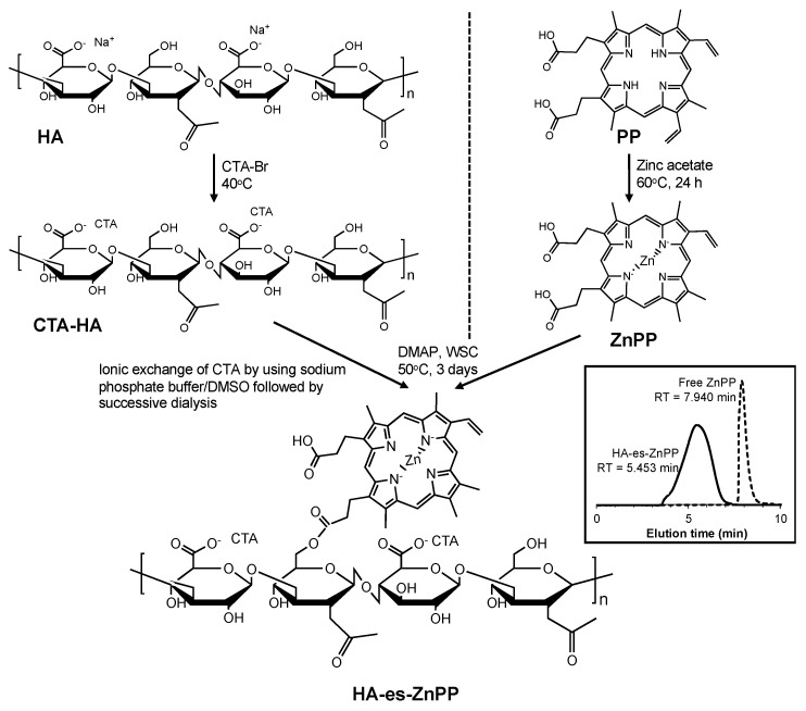
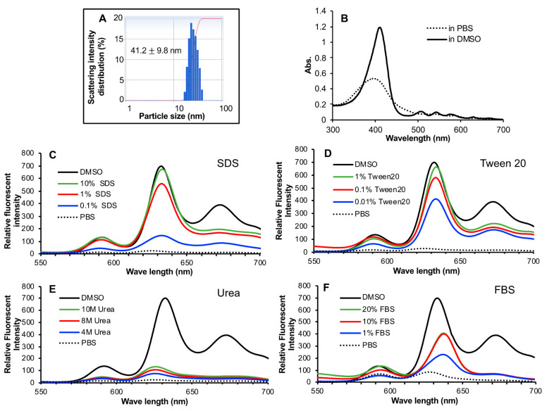
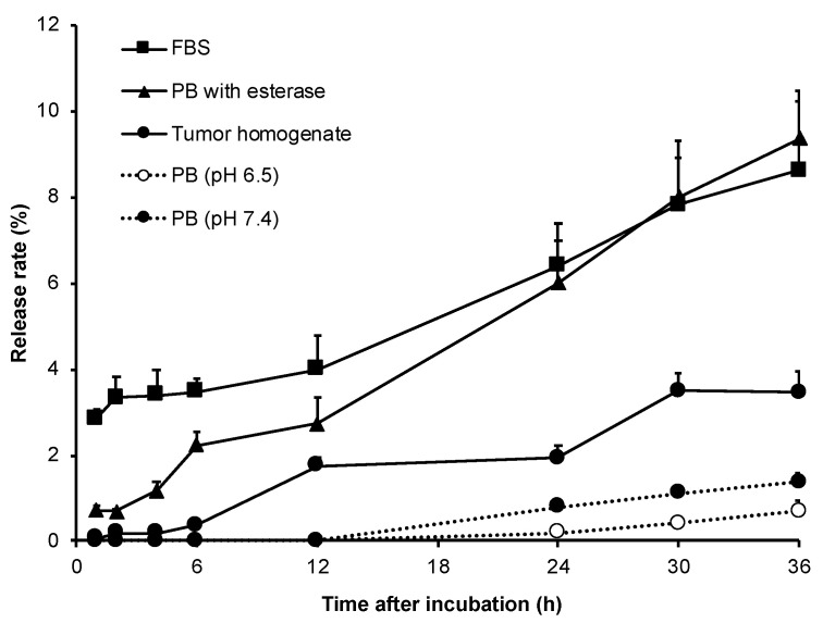
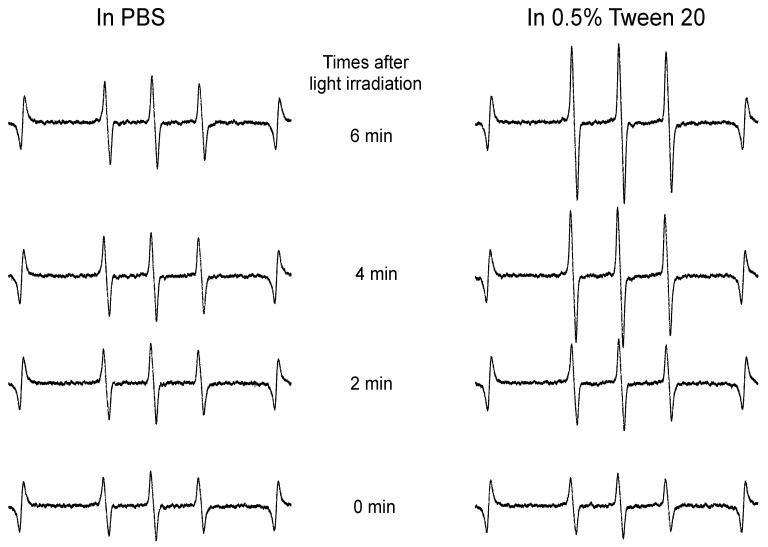
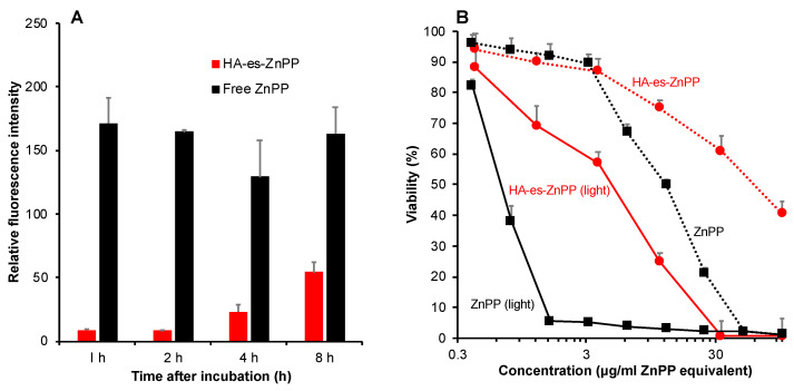
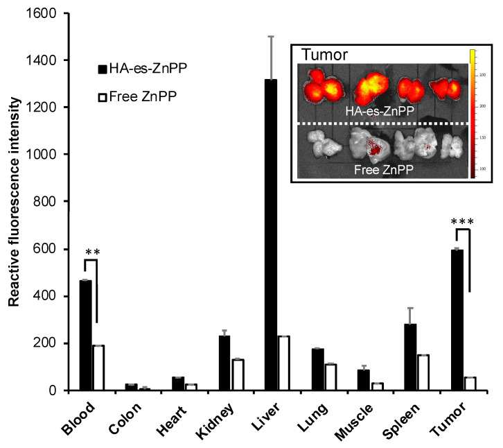
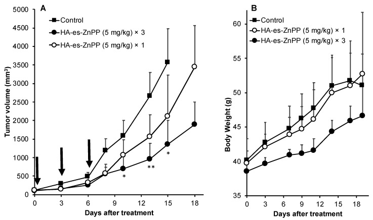
Targeted tumor accumulation, tumor environment responsive drug release, and effective internalization are critical issues being considered in developing anticancer nanomedicine. In this context, we synthesized a tumor environment-responsive nanoprobe for anticancer photodynamic therapy (PDT) that is a hyaluronan conjugated zinc protoporphyrin via an ester bond (HA-es-ZnPP), and we examined its anticancer PDT effect both in vitro and in vivo. HA-es-ZnPP exhibits high water-solubility and forms micelles of ~40 nm in aqueous solutions. HA-es-ZnPP shows fluorescence quenching without apparent 1O2 generation under light irradiation because of micelle formation. However, 1O2 was extensively generated when the micelle is disrupted, and ZnPP is released. Compared to native ZnPP, HA-es-ZnPP showed lower but comparable intracellular uptake and cytotoxicity in cultured mouse C26 colon cancer cells; more importantly, light irradiation resulted in 10-time increased cytotoxicity, which is the PDT effect. In a mouse sarcoma S180 solid tumor model, HA-es-ZnPP as polymeric micelles exhibited a prolonged systemic circulation time and the consequent tumor-selective accumulation based on the enhanced permeability and retention (EPR) effect was evidenced. Consequently, a remarkable anticancer PDT effect was achieved using HA-es-ZnPP and a xenon light source, without apparent side effects. These findings suggest the potential of HA-es-ZnPP as a candidate anticancer nanomedicine for PDT.

Keywords: EPR effect; hyaluronan; photodynamic; tumor targeting; zinc protoporphyrin.


Conflict of interest statement

The authors declare no conflict of interest.

Figures

Figure 1
Figure 1
Synthesizing protocol of hyaluronan (HA) conjugated ZnPP through ester bond (HA-es-ZnPP). Inset shows the gel permeation chromatograph of HA-es-ZnPP and free ZnPP, by using a high-performance liquid chromatography (HPLC) system with an Asahipak GF-310 HQ column.
Figure 2
Figure 2
Micelle formation of HA-es-ZnPP. HA-es-ZnPP showed a hydrodynamic diameter in physiological saline of 41.2 nm, as measured by DLS (A). The micelle formation was supported by decreased absorbance as seen in UV/vis spectra (B), and more importantly, fluorescence quenching, the fluorescence intensity of HA-es-ZnPP in PBS was almost undetectable compared to that in DMSO (C,D). The micelle formation could be effectively disrupted by detergent SDS (C) and Tween 20 (D), as evidenced by increased fluorescence, but it could not be affected by urea (E). Increased fluorescence intensity/disruption of micelles was also found in the presence of FBS (F). See text for details.
Figure 3
Figure 3
ZnPP release from HA-es-ZnPP at different conditions. In sodium phosphate buffer, almost no ZnPP was released from the conjugate at both physiological pH and weak acidic pH up to 12 h. Much rapid release of ZnPP occurred in presence of either esterase or FBS having esterase activities. A relatively high level of ZnPP release was also found in tumor homogenate.
Figure 4
Figure 4
Electron Spin Resonance (ESR) spectra of HA-es-ZnPP in phosphate-buffered saline (PBS) in the presence/absence of Tween 20 under light irradiation. 1O2 generation from HA-es-ZnPP was captured by 4-oxo-TEMP, and triplet 4-oxo-TEMP signal as the indicator of 1O2 was detected by ESR spectra. In the presence of Tween 20, a relatively high level of 1O2 generation was observed in an irradiation time-dependent manner, whereas no 1O2 generation occurred in PBS without Tween 20, in which HA-es-ZnPP behaves as micelles.
Figure 5
Figure 5
Intracellular uptake (A) and in vitro cytotoxicity (B) of HA-es-ZnPP in C26 colon cancer cells. (A), free ZnPP or HA-es-ZnPP (20 µg/mL ZnPP equivalent) was added to C26 cells for the indicated time. The intracellular ZnPP or HA-es-ZnPP was detected and quantified by measuring fluorescence intensity. (B), free ZnPP or HA-es-ZnPP at different concentrations was added into the cells, and the cells were treated for 48 h. In a separate plate, cells were irradiated by using fluorescent blue light (1.0 J/cm2, 5 min irradiation) at 24 h after HA-es-ZnPP treatment followed by further 24 h incubation. The cell viability was then measured by MTT assay. Data are mean ± SD.
Figure 6
Figure 6
In vivo tissue distribution of HA-es-ZnPP in S180 solid tumor-bearing ddY mice. HA-es-ZnPP or free ZnPP was injected i.v. in the mice. After 24 h, mice were killed, and tissues, including tumors were collected. Tissue samples were homogenated using DMSO, and extracted HA-es-ZnPP or ZnPP in the supernatant was quantified by fluorescence spectroscopy. Inset shows the fluorescence images of mice-tumors treated with free ZnPP or HA-es-ZnPP. The tumors were cut in the middle of tumor nodules, and the cross-sectional views were shown. Data are mean ± SD. **, p < 0.01; ***, p < 0.001.
Figure 7
Figure 7
In vivo therapeutic effect of HA-es-ZnPP based PDT. In a mouse sarcoma S180 solid tumor model, HA-es-ZnPP (5 mg/kg, ZnPP equivalent) was injected i.v. when the tumor grew to 8–10 mm in diameter. At 24 and 48 h after injection of HA-es-ZnPP, irradiation to the tumors was carried out using xenon light (MAX-303; Asahi Spectra) at 400–700 nm for 5 min (36 J/cm2). This treatment protocol was carried out 1 time or 3 times in different groups. The tumor volume (A) and body weight (B) of mice were recorded every 2–3 days during the study period. Arrows indicate the PDT treatments. Data are mean ± SD. *, p < 0.05; **, p < 0.01.

References

    1. Vogelstein B., Papadopoulos N. Cancer genome landscapes. Science. 2013;339:1546–1558. doi: 10.1126/science.1235122. - DOI - PMC - PubMed
    1. Gerlinger M., Rowan A.J. Intratumor heterogeneity and branched evolution revealed by multiregion sequencing. N. Engl. J. Med. 2012;366:883–892. doi: 10.1056/NEJMoa1113205. - DOI - PMC - PubMed
    1. Cutler D.M. Early returns from the era of precision medicine. JAMA. 2020;323:109–110. doi: 10.1001/jama.2019.20659. - DOI - PubMed
    1. Germain M., Caputo F. Delivering the power of nanomedicine to patients today. J. Control. Release. 2020;326:164–171. doi: 10.1016/j.jconrel.2020.07.007. - DOI - PMC - PubMed
    1. Lammers T., Ferrari M. The success of nanomedicine. Nano Today. 2020;31:100853. doi: 10.1016/j.nantod.2020.100853. - DOI - PMC - PubMed

LinkOut - more resources