Review
. 2021 Feb 17;11(2):331.
doi: 10.3390/diagnostics11020331.

Pyonephrosis Ultrasound and Computed Tomography Features: A Pictorial Review

Affiliations
Review

Pyonephrosis Ultrasound and Computed Tomography Features: A Pictorial Review

Stefania Tamburrini et al. Diagnostics (Basel). .

Abstract

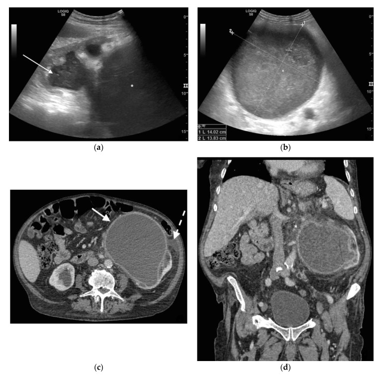
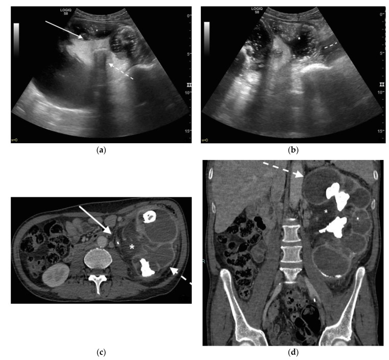
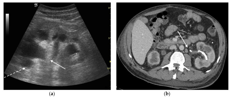
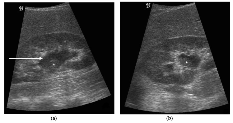
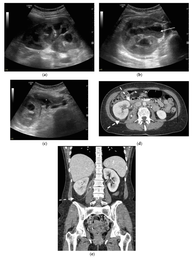
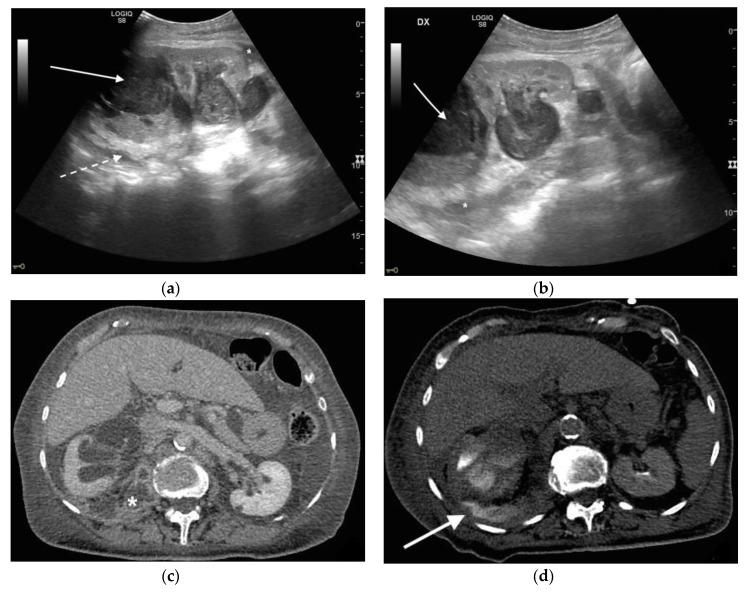
Urinary tract infections (UTIs) are the most frequent community-acquired and healthcare-associated bacterial infections. UTIs are heterogeneous and range from rather benign, uncomplicated infections to complicated UTIs (cUTIs), pyelonephritis and severe urosepsis, depending mostly on the host response. Ultrasound and computed tomography represent the imaging processes of choice in the diagnosis and staging of the pathology in emergency settings. The aim of this study is to describe the common ultrasound (US) and computed tomography (CT) features of pyonephrosis. US can make the diagnosis, demonstrating echogenic debris, fluid/fluid levels, and air in the collecting system. Although the diagnosis appears to be easily made with US, CT is necessary in non-diagnostic US examinations to confirm the diagnosis, to demonstrate the cause and moreover to stage the pathology, defining extrarenal complications. In emergency settings, US and CT are differently used in the diagnosis and staging of pyonephrosis.

Keywords: CT; US; pyonephrosis; sepsis; urinary tract infections.


Conflict of interest statement

The authors declare no conflict of interest.

Figures

Figure 1
Figure 1
A 78 years old septic male patient. Ultrasound (US) longitudinal (a) and axial (b) view of the left kidney showing high-grade hydronephrosis with gross dilatation of the renal pelvis and calyces, filled by inhomogeneous urine (white arrow) and an extremely dilated inferior calyceal group filled by a huge ball of debris conglomerate (*). Axial (c) and MPR coronal Computed Tomography (CT) (d) after intravenous contrast in cortical-medullary phase, showing high grade hydronephrosis, diffuse parietal thickening of the calico-pelvic system (white arrow), fat stranding of perirenal and renal sinus fat. Small amount of free fluid is appreciated anteriorly (dashed arrow).
Figure 2
Figure 2
A 71 years old septic patient. US longitudinal (a,b) view of left kidney. Severe hydronephrosis, with extreme cortical thinning. High-grade dilatation and parietal thickening (white arrow) of calico-pelvic system fluid filled by inhomogeneous fluid with multiple hyperechogenic spots (*). Enlarged and markedly hyperechogenicity of renal sinus fat with acoustic shadowing due to staghorn calculi (dashed arrow). Perirenal fat inhomogeneity was detected (dashed line). Axial (c) and MPR coronal (d) CT with intravenous contrast in cortical-medullary phase after stent placement (white arrow), showing high-grade hydronephrosis with diffuse parietal thickening of calico-pelvic system (dashed arrow), staghorn calculi and renal sinus lipomatosis with mild inhomogeneous fat stranding and suffusion (*).
Figure 3
Figure 3
A 73 years old septic male patient. US longitudinal view of left kidney (a,b) showing hydronephrosis with parietal thickening of inferior calyceal group (*), dense peripheral echoes within the calyceal system (white arrow) with acoustic shadowing at low-gain indicating gas-forming infection, perirenal suffusion was detected (dashed arrow). Axial CT (b) with intravenous contrast in cortical-medullary phase after stent placement (white arrow), showing drainage of calico-pelvic system. Mild perirenal fat stranding (*).
Figure 4
Figure 4
A 42 years old septic female patient. US longitudinal view (a,b) of the right kidney showing low/mild-grade hydronephrosis (white arrow) with sharply defined urine debris level (*).
Figure 5
Figure 5
A 48 years old septic woman. US longitudinal (a) and axial (b,c) view of the right kidney. Enlargement of the kidney, hydronephrosis with sharply defined urine-debris level (white arrow). Thickening of perirenal fascia and markedly inhomogeneous echogenicity of perirenal fat suggesting extrarenal extension (*). The patient underwent nephrostomy in the emergency setting, and CT was performed after stent placement. Axial (d) and MPR coronal (e) CT with intravenous contrast in parenchymal phase, showing correct stent positioning (white arrow) and drainage of calyceal-pelvic system. The right kidney appeared enlarged with a delayed parenchymal phase, the calyceal-pelvic system was unstretched after nephrostomy placement. Right perirenal fascia was thickened posteriorly (dashed arrow) and extrarenal extension of the pathology was confirmed by the presence of an abscess in the right iliopsoas muscle (*).
Figure 6
Figure 6
An 83 years old septic woman. US longitudinal (a,b) view of the right kidney showing high-grade hydronephrosis with the calyceal-pelvic system completely occupied by inhomogeneous echogenic debris (with arrow). Perirenal fascia is thickened and perirenal fat is hyperechogenic and inhomogeneous (dashed arrow). Fluid over-collection is appreciated around the kidney (*), and free fluid was appreciated in the abdomen. Complicated pyonephrosis with extrarenal extension was diagnosed. CT with intravenous contrast. Axial CT after intravenous contrast in parenchymal (c) and urography phase (d), showing hydronephrosis with parietal thickening of calyceal-pelvic system, multiple loculated over fluid collection in the perirenal (white arrow) space and abscess in iliopsoas muscle (*). In the urography phase, extravasation of urine into extrarenal fluid collections due to abscessualization of the calyceal-pelvic system.

Similar articles

Cited by

References

    1. Flores-Mireles A.L., Walker J.N., Caparon M.G., Hultgren S.J. Urinary tract infections: Epidemiology, mechanisms of infection and treatment options. Nat. Rev. Microbiol. 2015;13:269–284. doi: 10.1038/nrmicro3432. - DOI - PMC - PubMed
    1. Wagenlehner F.M.E., Johansen T.E.B., Cai T., Koves B., Kranz J., Pilatz A., Tandogdu Z. Epidemiology, definition and treatment of complicated urinary tract infections. Nat. Rev. Urol. 2020;17:586–600. doi: 10.1038/s41585-020-0362-4. - DOI - PubMed
    1. Wagenlehner F.M.E., Pilatz A., Weidner W., Naber K.G. Urosepsis: Overview of the Diagnostic and Treatment Challenges. Microbiol. Spectr. 2015;3 doi: 10.1128/microbiolspec.UTI-0003-2012. - DOI - PubMed
    1. Wang X., Tang K., Xia D., Peng E., Li R., Liu H., Chen Z. A novel comprehensive predictive model for obstructive pyonephrosis patients with upper urinary tract stones. Int. J. Clin. Exp. Pathol. 2020;13:2758–2766. - PMC - PubMed
    1. Patodia M., Goel A., Singh V., Singh B.P., Sinha R.J., Kumar M., Dalela D., Sankhwar S.N. Are there any predictors of pyonephrosis in patients with renal calculus disease? Urolithiasis. 2017;45:415–420. doi: 10.1007/s00240-016-0933-2. - DOI - PubMed

LinkOut - more resources