. 2021 Feb 17;22(4):1991.
doi: 10.3390/ijms22041991.

Enhanced Production of the Mical Redox Domain for Enzymology and F-actin Disassembly Assays

Affiliations

Enhanced Production of the Mical Redox Domain for Enzymology and F-actin Disassembly Assays

Jimok Yoon et al. Int J Mol Sci. .

Abstract

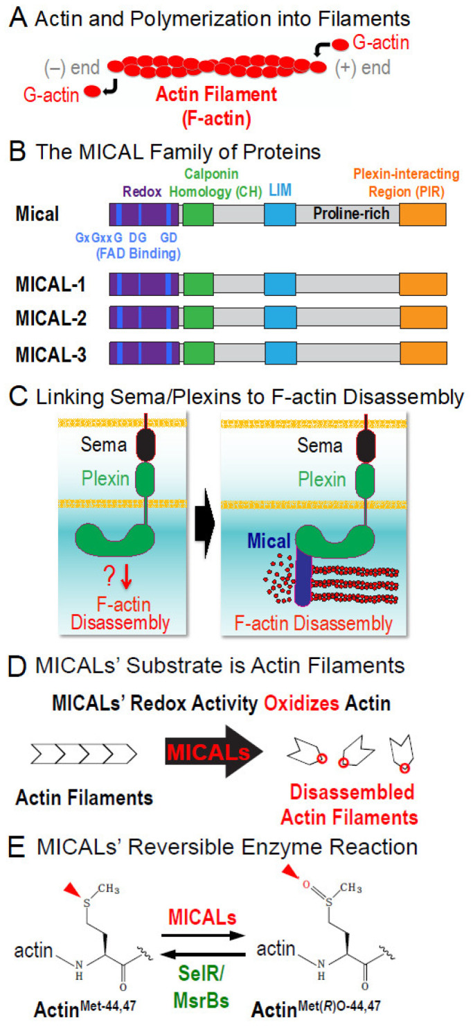
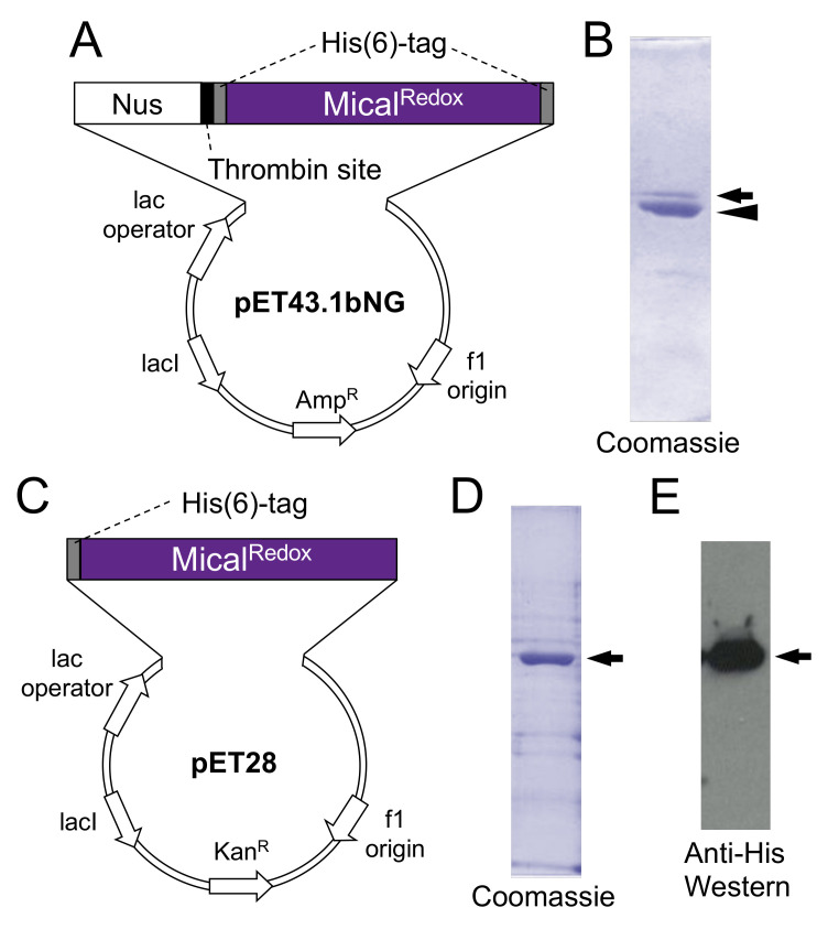
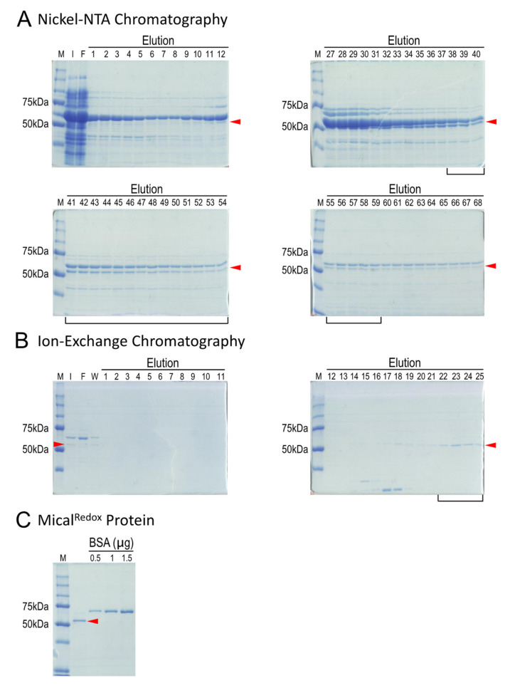
To change their behaviors, cells require actin proteins to assemble together into long polymers/filaments-and so a critical goal is to understand the factors that control this actin filament (F-actin) assembly and stability. We have identified a family of unusual actin regulators, the MICALs, which are flavoprotein monooxygenase/hydroxylase enzymes that associate with flavin adenine dinucleotide (FAD) and use the co-enzyme nicotinamide adenine dinucleotide phosphate (NADPH) in Redox reactions. F-actin is a specific substrate for these MICAL Redox enzymes, which oxidize specific amino acids within actin to destabilize actin filaments. Furthermore, this MICAL-catalyzed reaction is reversed by another family of Redox enzymes (SelR/MsrB enzymes)-thereby revealing a reversible Redox signaling process and biochemical mechanism regulating actin dynamics. Interestingly, in addition to the MICALs' Redox enzymatic portion through which MICALs covalently modify and affect actin, MICALs have multiple other domains. Less is known about the roles of these other MICAL domains. Here we provide approaches for obtaining high levels of recombinant protein for the Redox only portion of Mical and demonstrate its catalytic and F-actin disassembly activity. These results provide a ground state for future work aimed at defining the role of the other domains of Mical - including characterizing their effects on Mical's Redox enzymatic and F-actin disassembly activity.

Keywords: Drosophila; F-actin disassembly; MICAL1; MICAL2; MICAL3; oxidoreductase; plexin; repellent; repulsion; semaphorin.


Conflict of interest statement

The authors declare no conflict of interest. The funders had no role in the design of the study; in the collection, analyses, or interpretation of data; in the writing of the manuscript, or in the decision to publish the results.

Figures

Figure 1
Figure 1
The MICALs are a family of F-actin modifying and dismantling enzymes. (A) Single actin proteins (G-actin) form filaments (F-actin) to control numerous cellular behaviors and tissue functions. (B) The MICAL family of proteins are oxidoreductase enzymes. MICALs are conserved from invertebrates (e.g., Drosophila Mical) to vertebrates (MICAL-1, MICAL-2 and MICAL-3, which are coded for by 3 different genes). In addition to their Redox enzymatic region (purple), which have 3 conserved motifs for binding flavin adenine dinucleotide (FAD) (blue), MICALs have multiple other domains including a calponin homology (CH) domain (green), a LIM domain (cyan), a stretch of proline residues that serve as ligands for SH3-domain containing proteins (proline-rich) and a coiled-coil C-terminal region that binds the Semaphorin receptor Plexin (Plexin-interacting Region, orange), as well as Rab family small GTPases. (C) MICALs provide a means by which one of the largest protein families of extracellular cues, the Semaphorins (Sema) and their Plexin receptors, disassemble F-actin. (D) F-actin serves as a substrate for MICALs—such that the MICALs are activated by F-actin and then MICALs oxidize and disassemble actin filaments. In particular, Mical oxidizes the methionine (Met)-44 and 47 residues of actin (to form Met-44,47-sulfoxide). Met44 & 47 are located at the pointed end of actin where monomers (subunits) join to form a filament and their oxidation by Mical induces F-actin structural changes that trigger them to disassemble [13,14,15,16,19,20,25]. (E) Summary of Mical’s enzymatic reaction showing that individual actin subunits become stereospecifically (R) oxidized (O) on their Met44 & 47 residues by Mical (forming ActinMet(R)O-44,47). These effects of Mical on actin are reversed by SelR/MsrB methionine sulfoxide reductases.
Figure 2
Figure 2
Expression and purification of MicalRedox protein with different fusion partners and chaperonins. (A,B) Nus solubility tag and low-temperature chaperonins only allow expression and solubility of small amounts of the Redox only portion of Mical. (A) Vector (pET43.1bNG) and insert for expressing a Nus solubility tag fused to MicalRedox protein. (B) Following thrombin digestion to remove the Nus solubility tag, the expected 1:1 ratio of Nus:MicalRedox protein is affected. In particular, while the Nus tag remains soluble (arrowhead), little MicalRedox protein remains in the soluble fraction (arrow). (CE) Low-temperature chaperonins in combination with no solubility tags allow high expression and solubility of the Redox only portion of Mical. (C) Vector (pET28) and insert for expressing MicalRedox protein without a solubility tag. (D,E) High levels of MicalRedox protein is expressed and is soluble (arrows) with this expression strategy, as can be seen by Coomassie staining (D) and His antibody Western analysis (E).
Figure 3
Figure 3
Purification of recombinant MicalRedox protein. Coomassie stained bands are shown and the arrowheads point to the His(6)-tagged MicalRedox protein in all gels. (A) MicalRedox protein (arrowhead) eluted from Nickel columns was observed in multiple fractions. Cpn60 can also be seen (large band above the MicalRedox protein). The fractions containing MicalRedox protein (e.g., 11–68) were combined to load on to an ion-exchange column. In this particular purification, only fractions 38–59 (black bracket) were combined and used for loading on the ion-exchange column. (B) A strong cation exchange Mono S column was used to separate MicalRedox protein (arrowhead) from other contaminating proteins including Cpn60. Fractions enriched with MicalRedox protein (22–25; black bracket) were combined and the buffer system was changed to the storage buffer. (C) MicalRedox protein (arrowhead) and purity were checked by running on a gel next to known concentrations of bovine serum albumin (BSA) protein. M, protein markers; I, input; F, flow-through; W, wash.
Figure 4
Figure 4
Analysis of the activity of purified MicalRedox protein. (A) Mical’s enzymatic reaction with F-actin. This model is based on our results (reviewed in [9,10,11,12]) that Mical (1) physically associates with its substrate F-actin (2), which triggers Mical’s enzymatic activity to convert its co-enzyme nicotinamide adenine dinucleotide phosphate (NADPH) to NADP+ (3). Mical then oxidizes (Ox) F-actin subunits on their methionine (M) 44 and M47 residues (4), triggering F-actin disassembly. Note that for simplicity, details, such as the presence of molecular oxygen (O2) and conversion of flavin adenine dinucleotide (FAD) to FADH2 and reoxidation back FAD, have been left off the model. (B) Mical uses the pyridine nucleotide NADPH as a co-enzyme in its Redox enzymatic reactions [13,14,15,20]. We therefore sought to confirm the enzyme activity of the purified MicalRedox protein by characterizing its NADPH consumption activity. As judged by the decrease in absorbance over time (NADPH absorbs light at 340 nm, while NADP+ does not), our results revealed that our MicalRedox protein converts NADPH to NADP+ and therefore has enzyme activity. Both conditions contain NADPH. [NADPH] = 200 µM; [MicalRedox] = 50 nM; [F-actin] = 18.4 µM. (C,D) Pyrene-labeled actin depolymerization assays demonstrate purified MicalRedox protein’s ability to disassemble F-actin. (C) The fluorescence of pyrene-labeled actin is higher when actin is present in its polymerized form. (D) Similar to previous results with Mical [13,14,15,20], our newly purified MicalRedox protein induces actin depolymerization in the presence of its NADPH coenzyme as judged by a Pyrene-actin depolymerization assay, where the fluorescence of polymerized actin decreases as actin depolymerizes. [MicalRedox] = 50 nM; [NADPH] = 100 µM; [F-actin] = 4.65 µM.

Similar articles

Cited by

References

    1. Pollard T.D. Actin and Actin-Binding Proteins. Cold Spring Harb. Perspect. Biol. 2016;8:a018226. doi: 10.1101/cshperspect.a018226. - DOI - PMC - PubMed
    1. Goodson H.V., Jonasson E.M. Microtubules and Microtubule-Associated Proteins. Cold Spring Harb. Perspect. Biol. 2018;10:a022608. doi: 10.1101/cshperspect.a022608. - DOI - PMC - PubMed
    1. Terman J.R., Kashina A. Post-translational modification and regulation of actin. Curr. Opin. Cell Biol. 2013;25:30–38. doi: 10.1016/j.ceb.2012.10.009. - DOI - PMC - PubMed
    1. Varland S., Vandekerckhove J., Drazic A. Actin Post-translational Modifications: The Cinderella of Cytoskeletal Control. Trends Biochem. Sci. 2019;44:502–516. doi: 10.1016/j.tibs.2018.11.010. - DOI - PubMed
    1. Hung R.-J., Terman J.R. Extracellular inhibitors, repellents and Semaphorin/Plexin/MICAL-mediated actin filament disassembly. Cytoskeleton. 2011;68:415–433. doi: 10.1002/cm.20527. - DOI - PMC - PubMed

LinkOut - more resources