

MR-Imaging and Histopathological Diagnostic Work-Up of Patients with Spontaneous Lobar Intracerebral Hemorrhage: Results of an Institutional Prospective Registry Study
- PMID: 33671532
- PMCID: PMC7926429
- DOI: 10.3390/diagnostics11020368
MR-Imaging and Histopathological Diagnostic Work-Up of Patients with Spontaneous Lobar Intracerebral Hemorrhage: Results of an Institutional Prospective Registry Study
Abstract
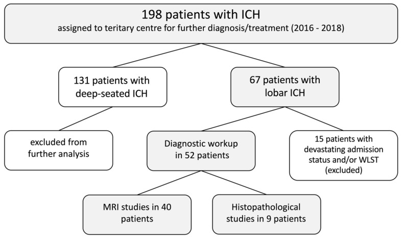
Intracerebral hemorrhage (ICH) is a frequently disabling or fatal disease. The localization of ICH often allows an etiological association. However, in atypical/lobar ICH, the cause of bleeding is less obvious. Therefore, we present prospective histopathological and radiological studies which were conducted within the diagnostic workup to identify causes for lobar ICH other than hypertension. From 2016 to 2018, 198 patients with spontaneous, non-traumatic ICH requiring neurosurgical monitoring were enrolled in an institutional prospective patient registry. Patients with deep-seated ICH and/or hemorrhagically transformed cerebral infarcts were excluded from further analysis. Data to evaluate the source of bleeding based on histopathological and/or radiological workup were prospectively evaluated and analyzed. After applying the inclusion criteria and excluding patients with incomplete diagnostic workup, a total of 52 consecutive patients with lobar ICH were further analyzed. Macrovascular disease was detected in 14 patients with lobar ICH (27%). In 11 patients, diagnostic workup identified cerebral amyloid angiopathy-related ICH (21%). In addition, five patients with tumor-related ICH (10%) and six patients with ICH based on infectious pathologies (11%) were identified. In four patients, the cause of bleeding remained unknown despite extensive diagnostic workup (8%). The present prospective registry study demonstrates a higher probability to identify a cause of bleeding other than hypertension in patients with lobar ICH. Therefore, a thorough diagnostic work-up in patients with ICH is essential to accelerate treatment and further improve outcome or prevent rebleeding.
Keywords: AVM; CAA; diagnostic work-up; etiology; intracerebral hemorrhage; tumor.
Conflict of interest statement
The authors declare no conflict of interest. The funders had no role in the design of the study; in the collection, analyses, or interpretation of data; in the writing of the manuscript, or in the decision to publish the results.
Figures
References
-
- Kitchen P., Salman M.M., Halsey A.M., Clarke-Bland C., Macdonald J.A., Ishida H., Vogel H.J., Almutiri S., Logan A., Kreida S., et al. Targeting Aquaporin-4 Subcellular Localization to Treat Central Nervous System Edema. Cell. 2020;181:784–799.e19. doi: 10.1016/j.cell.2020.03.037. - DOI - PMC - PubMed
Grants and funding
LinkOut - more resources
Full Text Sources
Other Literature Sources
