Review
. 2021 Feb 22;10(2):464.
doi: 10.3390/cells10020464.

Compartmentalized Signaling in Aging and Neurodegeneration

Affiliations
Review

Compartmentalized Signaling in Aging and Neurodegeneration

Giulietta Di Benedetto et al. Cells. .

Abstract

The cyclic AMP (cAMP) signalling cascade is necessary for cell homeostasis and plays important roles in many processes. This is particularly relevant during ageing and age-related diseases, where drastic changes, generally decreases, in cAMP levels have been associated with the progressive decline in overall cell function and, eventually, the loss of cellular integrity. The functional relevance of reduced cAMP is clearly supported by the finding that increases in cAMP levels can reverse some of the effects of ageing. Nevertheless, despite these observations, the molecular mechanisms underlying the dysregulation of cAMP signalling in ageing are not well understood. Compartmentalization is widely accepted as the modality through which cAMP achieves its functional specificity; therefore, it is important to understand whether and how this mechanism is affected during ageing and to define which is its contribution to this process. Several animal models demonstrate the importance of specific cAMP signalling components in ageing, however, how age-related changes in each of these elements affect the compartmentalization of the cAMP pathway is largely unknown. In this review, we explore the connection of single components of the cAMP signalling cascade to ageing and age-related diseases whilst elaborating the literature in the context of cAMP signalling compartmentalization.

Keywords: PKA; aging; cAMP; compartmentalization; neurodegeneration.


Conflict of interest statement

The authors declare no conflict of interest. The funders had no role in the design of the study; in the collection, analyses or interpretation of data; in the writing of the manuscript; or in the decision to publish the results.

Figures

Figure 1
Figure 1
Compartmentalization mechanisms of the cAMP/PKA axis. Cyclic AMP can be produced at the plasma membrane by transmembrane adenylyl cyclases (tmACs) or intracellularly, by the soluble adenylyl cyclase (sAC). Once produced, the levels of cAMP are regulated by 3 main mechanisms: degradation by phosphodiesterases (PDEs), extrusion from the cells to the extracellular milieu by multi drug resistance channels (MDR channels) or buffering by RI alpha-constituted membraneless organelles. A final, cAMP-independent, regulatory step is exerted by the action of phosphatases (PPs) that by dephosphorylating PKA-phosphorylated targets effectively terminate the cascade. Created with BioRender.com.
Figure 2
Figure 2
Schematic representation of the main AC5-driven pathways in ageing. (left panel): AC5 overexpression leads to enhanced oxidative stress through the SIRT1/FoxO3a axis. Conversely, disruption of AC5 leads to beneficial effects through the SIRT1/FoxO3 activity and the Raf/MEK/ERK signalling cascade, both impinging on the MnSOD levels (right panel). Created with BioRender.com.
Figure 3
Figure 3
AKAP79/150 is involved in the coordination of synaptic plasticity. At the PSD, AKAP79/150 assembles a multiproteic complex containing β2-ARs, AMPARs, PKA, CaN and an adenylyl cyclase (likely AC5). This complex is involved in NMDAR-independent LTP, where stimulation of β2-ARs (by norepinephrine, NE) results in increased PKA dependent phosphorylation of AMPAR GluR1 and of CaV1.2 LTCCs. PKA phosphorylation promotes exocytosis, lateral diffusion and synaptic accumulation of AMPARs, and increases the opening probability of both AMPAR and LTCCs. Activation of CaN by Ca2+ influx through LTCC counteracts phosphorylation of both LTCC, as a negative feedback regulation to hinder Ca2+ entry, and AMPAR. Dephosphorylated AMPAR are removed from the synapse (not shown), leading to LTD. Moreover, activation of CaN leads to the dephosphorylation of NFAT, promoting its translocation in the nucleus, where it can act as transcriptional activator/repressor. Persistent CaN activation leads to down-regulation of NFAT targets including synaptic genes, resembling the transcriptional profiles encountered in human AD brain. A similar complex assembled by AKAP79/150 close to NMDAR (not shown) mediates LTP and contains AMPAR, PKA, CN and a Ca2+-activated AC (likely AC1 or AC8). Created with BioRender.com.

References

    1. Nikolaev V.O., Moshkov A., Lyon A.R., Miragoli M., Novak P., Paur H., Lohse M.J., Korchev Y.E., Harding S.E., Gorelik J. 2-Adrenergic Receptor Redistribution in Heart Failure Changes cAMP Compartmentation. Science. 2010;327:1653–1657. doi: 10.1126/science.1185988. - DOI - PubMed
    1. Baillie G.S., Tejeda G.S., Kelly M.P. Therapeutic targeting of 3′,5′-cyclic nucleotide phosphodiesterases: Inhibition and beyond. Nat. Rev. Drug Discov. 2019;18:770–796. doi: 10.1038/s41573-019-0033-4. - DOI - PMC - PubMed
    1. Torres-Quesada O., Mayrhofer J.E., Stefan E. The many faces of compartmentalized PKA signalosomes. Cell. Signal. 2017;37:1–11. doi: 10.1016/j.cellsig.2017.05.012. - DOI - PubMed
    1. Grisan F., Burdyga A., Iannucci L.F., Surdo N.C., Pozzan T., Di Benedetto G., Lefkimmiatis K. Studying β1 and β2 adrenergic receptor signals in cardiac cells using FRET-based sensors. Prog. Biophys. Mol. Biol. 2019 doi: 10.1016/j.pbiomolbio.2019.06.001. - DOI - PubMed
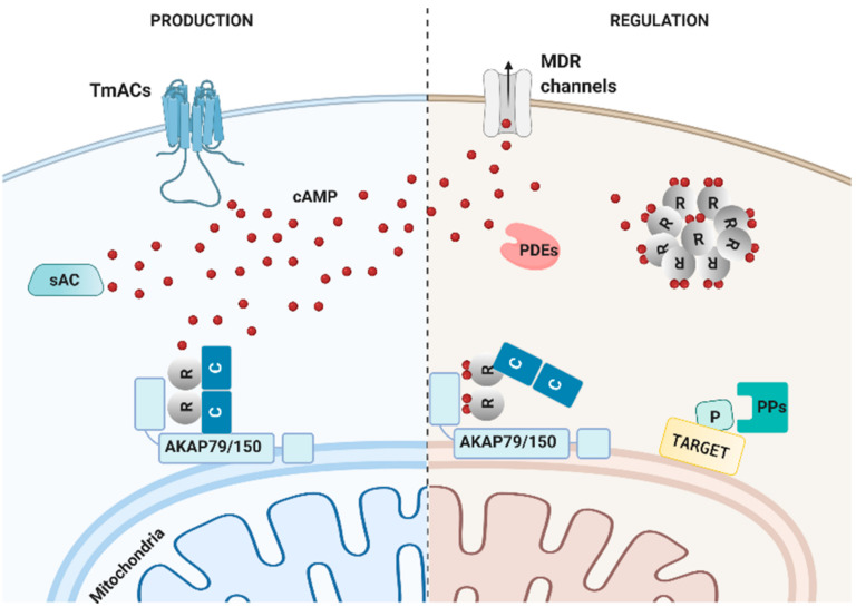
    1. Burdyga A., Surdo N.C., Monterisi S., Di Benedetto G., Grisan F., Penna E., Pellegrini L., Zaccolo M., Bortolozzi M., Swietach P., et al. Phosphatases control PKA-dependent functional microdomains at the outer mitochondrial membrane. Proc. Natl. Acad. Sci. USA. 2018;115:E6497–E6506. doi: 10.1073/pnas.1806318115. - DOI - PMC - PubMed

Publication types

LinkOut - more resources