Review
. 2021 Feb 22;10(2):465.
doi: 10.3390/cells10020465.

Mechanical Mechanisms of Chromosome Segregation

Affiliations
Review

Mechanical Mechanisms of Chromosome Segregation

Maya I Anjur-Dietrich et al. Cells. .

Abstract

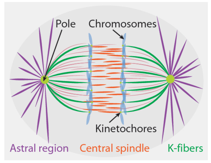
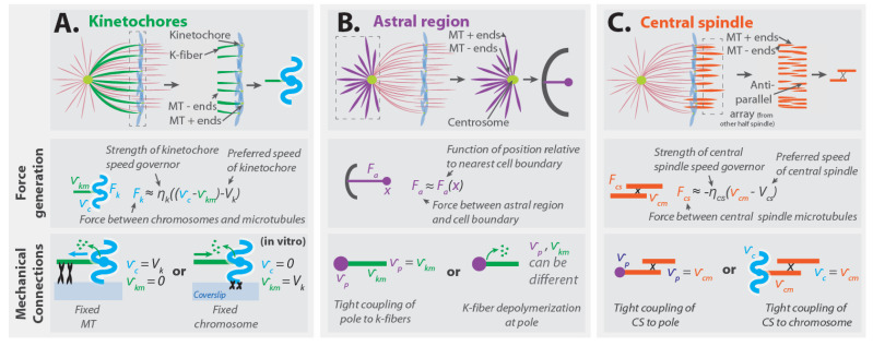
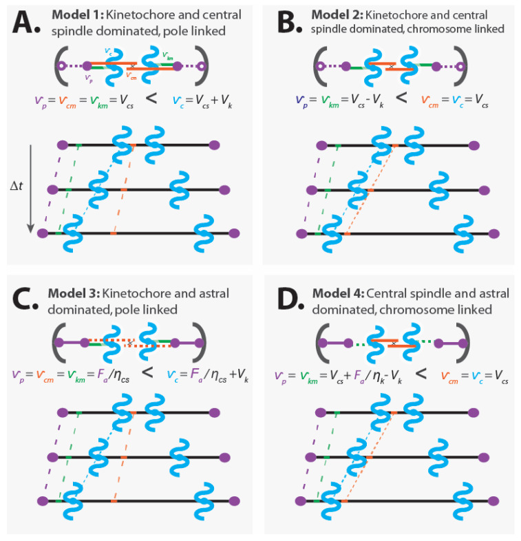
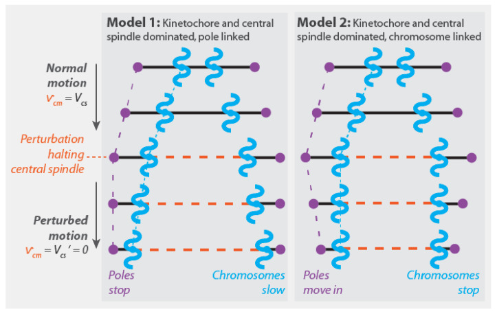
Chromosome segregation-the partitioning of genetic material into two daughter cells-is one of the most crucial processes in cell division. In all Eukaryotes, chromosome segregation is driven by the spindle, a microtubule-based, self-organizing subcellular structure. Extensive research performed over the past 150 years has identified numerous commonalities and contrasts between spindles in different systems. In this review, we use simple coarse-grained models to organize and integrate previous studies of chromosome segregation. We discuss sites of force generation in spindles and fundamental mechanical principles that any understanding of chromosome segregation must be based upon. We argue that conserved sites of force generation may interact differently in different spindles, leading to distinct mechanical mechanisms of chromosome segregation. We suggest experiments to determine which mechanical mechanism is operative in a particular spindle under study. Finally, we propose that combining biophysical experiments, coarse-grained theories, and evolutionary genetics will be a productive approach to enhance our understanding of chromosome segregation in the future.

Keywords: anaphase; chromosome segregation; mechanics; spindle.


Conflict of interest statement

The authors declare no conflict of interest.

Figures

Figure 1
Figure 1
Three force-generating sites that are found in diverse spindles are kinetochores, the astral region, and the central spindle.
Figure 2
Figure 2
The motion of an isolated chromosome dragged by an optical tweezer. The velocity of the chromosome is determined by the balance of the applied force from the optical tweezer, Fapplied, and the fluid drag force, Fdrag=ηcytov.
Figure 3
Figure 3
(A) Nicklas used a glass needle to apply force on anaphase chromosomes and slowed their motion. (B) He characterized the relationship between the applied force and velocity using a force–velocity curve. When no force is applied, the chromosomes move at the spindle’s preferred speed. Since the spindle has a preferred speed, it contains a speed governor. The stall force is the applied force that causes the chromosomes to stop moving. The origin of the speed governor in the spindle remains unclear, and, as will be elaborated on in Section 4, it might not reflect the force-generating properties of the kinetochore.
Figure 4
Figure 4
((A), top) A coarse-grained mechanical model of kinetochore forces and K-fibers, which are bundles composed of kinetochore microtubules. ((A), middle) Kinetochores apply forces between chromosomes and kinetochore microtubules, Fk. This force depends on the extent to which the relative velocity of chromosomes, vc, and kinetochore microtubules, vkm, differs from the kinetochore’s preferred speed, Vk. ((A), bottom) In vitro movement of the microtubules and chromosomes is dependent on which mechanical linkages are present. ((B), top) A coarse-grained mechanical model of the astral region forces. ((B), middle) The astral region applies forces between the pole and the cell boundary, Fa. This force depends on the distance between the pole and the cell boundary, x. ((B), bottom) Two different possible couplings with implications for the relationship between the velocity of kinetochore microtubules, vkm, and the velocity of spindle poles, vp. ((C), top) A coarse-grained mechanical model of central spindle forces. ((C), middle) The central spindle applies a force between the central spindle microtubules, Fcs. This force depends on the extent to which the velocity of the central spindle microtubules, vcm, differs from the central spindle’s preferred speed, Vcs. ((C), bottom) Two different possible couplings of central spindle microtubules: either tightly coupled to chromosomes or tightly coupled to poles.
Figure 5
Figure 5
(A) In Model 1, the central spindle microtubules slide apart at their preferred speed, Vcs, the kinetochore causes the kinetochore microtubules and chromatids to move relative to each other at its preferred speed, Vk, and the central spindle microtubules are rigidly attached to the spindle poles. In this model, the speed that chromosomes move, vc, is faster than the speed that central spindle microtubules move, vcm (which is equal to the speed of spindle poles, vp, and kinetochore microtubules, vkm). (B) In Model 2, the central spindle microtubules slide apart at their preferred speed, the kinetochore causes the kinetochore microtubules and chromatids to move relative to each other at its preferred speed, and the central spindle microtubules are rigidly attached to the chromatids. In this model, chromosomes move at the same speed as central spindle microtubules. (C) In Model 3, the astral region generates substantial forces, Fa, that pull the spindles poles apart with speed Faηcs (where ηcs is a measure of the strength of frictional forces between central spindle microtubules), and the central spindle microtubules are rigidly attached to the pole and passively respond to the forces acting on them. The kinetochore causes the kinetochore microtubules and chromatids to move relative to each other at its preferred speed. In this model, chromosomes move faster than the central spindle microtubules. (D) In Model 4, the astral region generates substantial forces that are sufficient to perturb the dynamics of the kinetochore (where ηk is a measure of the strength of kinetochore force generation), while the central spindle microtubules slide apart at their preferred speed. The central spindle microtubules are rigidly attached to the chromatids. In this model, chromosomes move at the same speed as the central spindle microtubules.
Figure 6
Figure 6
Experimental perturbations can be used to differentiate between different mechanical models of anaphase. For a perturbation that selectively halts the motion of the central spindle microtubules (i.e., by causing the preferred speed of the central spindle microtubules, Vcs, to become zero, VcsVcs=0), Models 1 and 2 (Figure 5A,B) both predict that the speed of the central spindle microtubules, vcm, becomes zero (i.e., vcm=Vcs=0). However, this change leads to different subsequent motions of chromosomes and poles in the two models. In Model 1, this perturbation causes spindle poles to cease moving, while chromosome motion continues (at a reduced rate). In Model 2, this perturbation causes chromosomes to cease moving. In both Models 1 and 2, this perturbation is predicted to have no impact on the relative motion of chromosomes and poles.

References

    1. Weinberg R.A. The Biology of Cancer. 2nd illus. ed. Garland Science; New York, NY, USA: 2013.
    1. McIntosh J.R., Hays T. A Brief History of Research on Mitotic Mechanisms. Biology. 2016;5:55. doi: 10.3390/biology5040055. - DOI - PMC - PubMed
    1. Pavin N., Tolić I.M. Mechanobiology of the Mitotic Spindle. Dev. Cell. 2021;56:192–201. doi: 10.1016/j.devcel.2020.11.003. - DOI - PubMed
    1. Maiato H., Lince-Faria M. The perpetual movements of anaphase. Cell. Mol. Life Sci. 2010;67:2251–2269. doi: 10.1007/s00018-010-0327-5. - DOI - PMC - PubMed
    1. Civelekoglu-Scholey G., Scholey J.M. Mitotic force generators and chromosome segregation. Cell. Mol. Life Sci. 2010;67:2231–2250. doi: 10.1007/s00018-010-0326-6. - DOI - PMC - PubMed

Publication types

LinkOut - more resources