. 2021 Feb 22;22(4):2162.
doi: 10.3390/ijms22042162.

Integrin-Linked Kinase Links Integrin Activation to Invadopodia Function and Invasion via the p(T567)-Ezrin/NHERF1/NHE1 Pathway

Affiliations

Integrin-Linked Kinase Links Integrin Activation to Invadopodia Function and Invasion via the p(T567)-Ezrin/NHERF1/NHE1 Pathway

Maria Raffaella Greco et al. Int J Mol Sci. .

Abstract

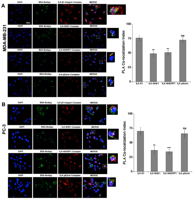
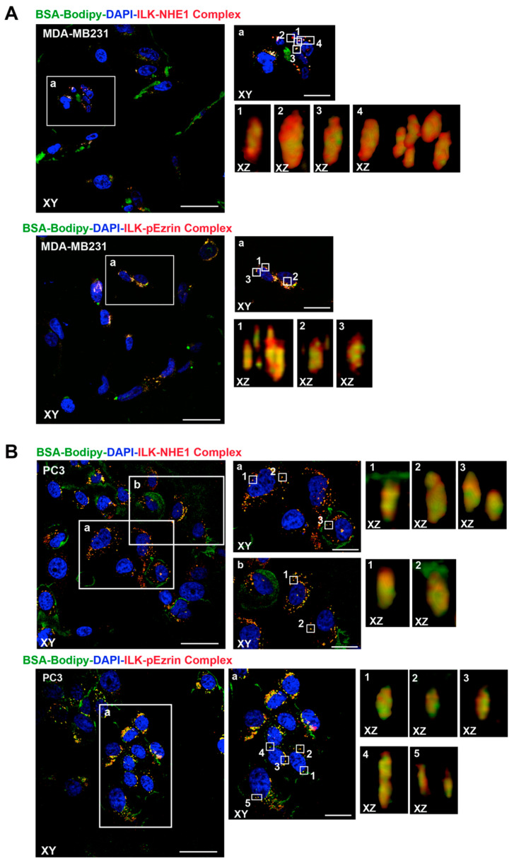
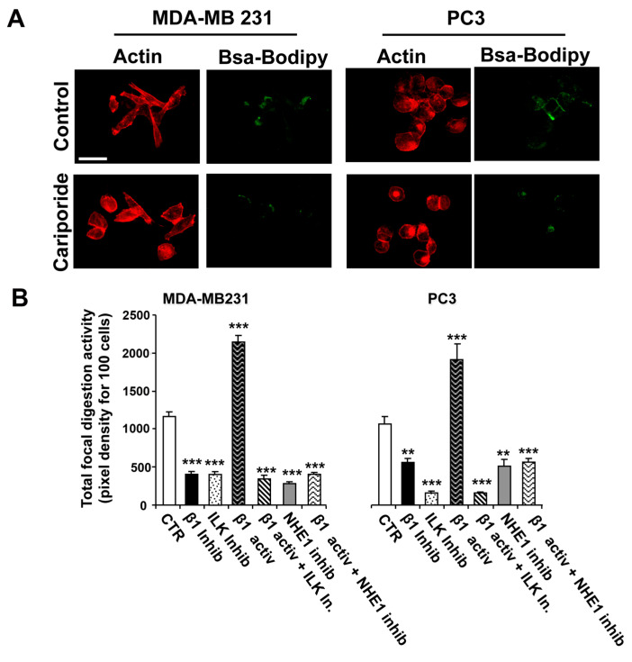
Tumor cell invasion depends largely on degradation of the extracellular matrix (ECM) by protease-rich structures called invadopodia, whose formation and activity requires the convergence of signaling pathways engaged in cell adhesion, actin assembly, membrane regulation and ECM proteolysis. It is known that β1-integrin stimulates invadopodia function through an invadopodial p(T567)-ezrin/NHERF1/NHE1 signal complex that regulates NHE1-driven invadopodia proteolytic activity and invasion. However, the link between β1-integrin and this signaling complex is unknown. In this study, in metastatic breast (MDA-MB-231) and prostate (PC-3) cancer cells, we report that integrin-linked kinase (ILK) integrates β1-integrin with this signaling complex to regulate invadopodia activity and invasion. Proximity ligation assay experiments demonstrate that, in invadopodia, ILK associates with β1-integrin, NHE1 and the scaffold proteins p(T567)-ezrin and NHERF1. Activation of β1-integrin increased both invasion and invadopodia activity, which were specifically blocked by inhibition of either NHE1 or ILK. We conclude that ILK integrates β1-integrin with the ECM proteolytic/invasion signal module to induce NHE1-driven invadopodial ECM proteolysis and cell invasion.

Keywords: breast cancer; integrin signaling; invadopodia; invasion; pH; prostate cancer.


Conflict of interest statement

The authors declare no conflict of interest.

Figures

Figure 1
Figure 1
ILK formed protein–protein complexes with β1-integrin receptor, p-ezrin, NHERF1 and NHE1 in areas of focal digestion of Matrigel in MDA-MB-231 (A) and PC-3 (B) cells. To better visualize invadopodial focal digestion and protein–protein complex localization in Matrigel, we utilized PLA for each protein–protein complex together with in situ zymography using the quenched fluorescent substrate, DQ Green-BSA. Therefore, quantifiable fluorescence was released only upon digestion of the matrix. After the cells digested the fluorogenic substrate (green), the cells were fixed for subsequent PLA analysis (red). The white arrows indicate areas of co-localization of BSA-Bodipy with the PLA signal. The histograms display the analysis of co-localization of ILK with the other proteins (PLA co-localization index) in the specific area of focal proteolysis in ECM digesting cells. Mean ± S.E.M., n = 6, ns: non-significant, ** p < 0.01, *** p < 0.001 for co-localization index compared to the ILK-β1 PLA analysis.
Figure 2
Figure 2
ILK formed protein–protein complexes with NHE1 and p-ezrin within invadopodia in MDA-MB-231 (A) and PC-3 (B) cells. Cells seeded on Matrigel were allowed to digest the green fluorogenic substrate (DQ) and PLA co-localization assays after fixation. Confocal images in axial planes taken at the bottom of the cells (XY) of a typical region showed protein–protein complexes (red) and digestion (green) localization. In each field, zoomed sections (XZ) reconstructed by alpha blending analysis of the indicated regions of interest (white box) are shown on the right. Importantly, protein–protein complexes (red) and digestion (green) were co-localized in protrusive digestive structures on the ventral cell surface. Scale bars = 10 µm (XY) and 5 µm (XZ).
Figure 3
Figure 3
ILK and NHE1 activity are necessary for β1-integrin-driven invadopodia proteolytic activity. To examine the role of NHE1 and ILK in invadopodial-dependent focal digestion of the ECM, MDA-MB-231 and PC3 cells were plated on Matrigel with DQ-Green BSA and, 1 h later, were treated with either the NHE1 inhibitor, ILK inhibitor, β1-integrin inhibiting antibody or β1-integrin activating antibody, with the β1-integrin activating antibody added in either the presence or the absence of the specific NHE1 or ILK inhibitor. After 24 h, ECM digestion was analyzed using fluorescence microscopy for a series of individual cells as described in the Materials and Methods. (A) Typical experiment showing control and cariporide-treated cell. (B) Histograms showing Mean ± S.E.M., n = 4, ** p < 0.05, *** p < 0.001 for focal proteolysis compared to the control cells.
Figure 4
Figure 4
ILK and NHE1 activity are necessary for β1-integrin-driven invasion. To examine the roles of ILK and NHE1 in an invasive capacity, MDA-MB-231 and PC3 cells were stimulated with the β1 integrin activating antibody and treated with either the ILK inhibitor or the β1 integrin inhibiting antibody in the presence or absence of the specific NHE1 inhibitor, cariporide. Cell invasion was analyzed quantitatively by fluorescent labeling of cells that had traversed 8 µm polycarbonate membranes coated with 5 mg/mL Matrigel (Chemicon Int., Livermore, CA, USA) as described in the Materials and Methods. Mean ± S.E.M., n = 4, *** p < 0.001 compared to control cells.
Figure 5
Figure 5
Model of the localization and role of ILK in NHE1-driven invadopodia formation and function. The insert is a magnification of the cellular membrane extrusion, the invadopodia, into the ECM. Invadopodia are F-actin-enriched membrane protrusions responsible for ECM degradation, whose formation is activated by β1-integrin binding to the ECM. This results in β1-integrin binding to and its activation of ILK and in the phosphorylation of the adapter protein, ezrin, at threonine 567. P-ezrin binds to NHE1 and the cytoskeleton and shifts the complex to PIP2-rich lipid rafts where NHE1 is activated [25]. NHE1, with its two functions as a scaffolding protein and ion exchanger, leads to membrane protrusion and proteolysis. As a proton transporter, NHE1 promotes invasion through its control of the acidification of the peri-invadopodial space, where NHE1 proton-secreting activity and proteases act in concert to degrade the ECM during invasion. The proteases cathepsin B, D and L, urokinase plasmogen activator and the matrix metalloproteinases MMP-2 and MMP-9 are released extracellularly, while MT1-MMP is associated with the membrane and participates, together with cathepsin B, in the processing of inactive pro-MMP-2 into active MMP-2. Glycolytic enzymes are enriched in invadopodia, leading to the localized production of intracellular protons secreted via active NHE1, resulting in peri-invadopodial acidification favorable for the activity of the various proteases localized in this sub-cellular region. Furthermore, the NHE1-dependent alkalinization of the invadopodia cytosol results in phosphorylation of cortactin with subsequent release of cofilin, which promotes actin polymerization, growth of the invadopodia cytoskeleton and invadopodia protrusion. NHE1 also promotes invadopodial formation via its interaction with the cytoskeleton through binding to the actin-anchoring protein, phospho-ezrin.

References

    1. Welch D.R., Hurst D.R. Defining the Hallmarks of Metastasis. Cancer Res. 2019;79:3011–3027. doi: 10.1158/0008-5472.CAN-19-0458. - DOI - PMC - PubMed
    1. Valastyan S., Weinberg R.A. Tumor metastasis: Molecular insights and evolving paradigms. Cell. 2011;147:275–292. doi: 10.1016/j.cell.2011.09.024. - DOI - PMC - PubMed
    1. Lehembre F., Regenass U. Metastatic disease: A drug discovery perspective. Semin. Cancer Biol. 2012;22:261–271. doi: 10.1016/j.semcancer.2012.03.005. - DOI - PubMed
    1. Fidler I.J., Kripke M.L. The challenge of targeting metastasis. Cancer Metastasis Rev. 2015;34:635–641. doi: 10.1007/s10555-015-9586-9. - DOI - PMC - PubMed
    1. Lambert A.W., Pattabiraman D.R., Weinberg R.A. Emerging Biological Principles of Metastasis. Cell. 2017;168:670–691. doi: 10.1016/j.cell.2016.11.037. - DOI - PMC - PubMed

LinkOut - more resources