. 2021 Feb 22;11(2):373.
doi: 10.3390/diagnostics11020373.

Comparison of Lung Ultrasound versus Chest X-ray for Detection of Pulmonary Infiltrates in COVID-19

Affiliations

Comparison of Lung Ultrasound versus Chest X-ray for Detection of Pulmonary Infiltrates in COVID-19

María Mateos González et al. Diagnostics (Basel). .

Abstract

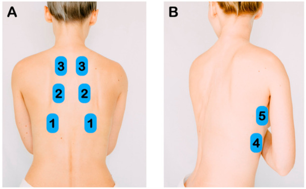
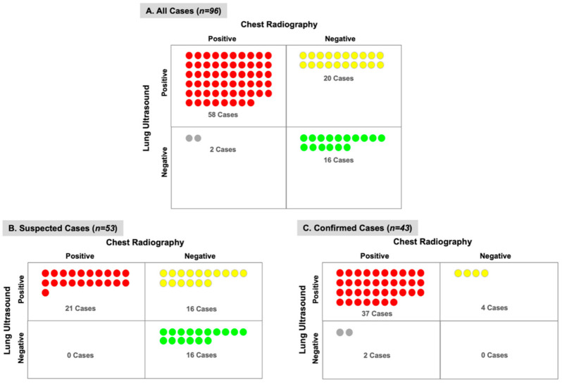
Point-of-care lung ultrasound (LUS) is an attractive alternative to chest X-ray (CXR), but its diagnostic accuracy compared to CXR has not been well studied in coronavirus disease 2019 (COVID-19) patients. We conducted a prospective observational study to assess the correlation between LUS and CXR findings in COVID-19 patients. Ninety-six patients with a clinical diagnosis of COVID-19 underwent an LUS exam and CXR upon presentation. Physicians blinded to the CXR findings performed all LUS exams. Detection of pulmonary infiltrates by CXR versus LUS was compared between patients categorized as suspected or confirmed COVID-19 based on reverse transcriptase-polymerase chain reaction. Sensitivities and correlation by Kappa statistic were calculated between LUS and CXR. LUS detected pulmonary infiltrates more often than CXR in both suspected and confirmed COVID-19 subjects. The most common LUS abnormalities were discrete B-lines, confluent B-lines, and small subpleural consolidations. Most important, LUS detected unilateral or bilateral pulmonary infiltrates in 55% of subjects with a normal CXR. Substantial agreement was demonstrated between LUS and CXR for normal, unilateral or bilateral findings (Κ = 0.48 (95% CI 0.34 to 0.63)). In patients with suspected or confirmed COVID-19, LUS detected pulmonary infiltrates more often than CXR, including more than half of the patients with a normal CXR.

Keywords: SARS; X-ray; chest; diagnosis; imaging; ultrasound.


Conflict of interest statement

The authors declare no conflict of interest. The funders had no role in the design of the study; in the collection, analyses, or interpretation of data; in the writing of the manuscript, or in the decision to publish the results.

Figures

Figure 1
Figure 1
Lung Ultrasound Exam Points. (A) After identifying the diaphragm, the transducer was slide cephalad to image the inferior, middle, and superior zones of the posterior chest. (B) Along the mid-axillary line, the inferior and superior lung zones of the lateral chest were imaged.
Figure 2
Figure 2
Characteristic Lung Lesions in coronavirus disease 2019 (COVID-19). (A) Normal lung ultrasound is defined by visualization of pleural sliding and A-lines. (B) Discrete B-lines are individual hyperechoic, laser-like artifacts the emanate from the pleural line and are due to increased interstitial fluid in the acute setting. Discrete B-lines are typically the first sign of COVID-19. (C) Fused or confluent B-lines are seen when individual B-lines coalesce as interstitial fluid increases. (D) Subpleural consolidations are typically small (<3 cm) areas of consolidation that are seen just below the pleural line.
Figure 3
Figure 3
Chest X-ray and Lung Ultrasound for Detection of Pulmonary Infiltrates. The number of suspected or confirmed COVID-19 subjects (n) with or without pulmonary infiltrates detected by chest X-ray or lung ultrasound is demonstrated. In both suspected and confirmed COVID-19 subjects, lung ultrasound was able to detect pulmonary infiltrates more often than chest radiography.
Figure 4
Figure 4
Correlation of Chest X-ray and Lung Ultrasound in Detection of Pulmonary Infiltrates. The number of subjects (n) and agreement between chest X-ray and lung ultrasound is shown for (A) all cases, (B) suspected COVID-19 cases, and (C) confirmed COVID-19 cases. Lung ultrasound detected pulmonary infiltrates in 20 subjects with a normal chest X-ray, whereas chest X-ray detected pulmonary infiltrates in 2 subjects with a normal LUS exam.
Figure 5
Figure 5
Distribution of Pulmonary Infiltrates Detected by Chest X-ray vs. Lung Ultrasound. The number of subjects (n) with confirmed or suspected COVID-19 who had pulmonary infiltrates detected in the upper, middle, or lower lobes of the right and left lung is demonstrated.

References

    1. Huang C., Wang Y., Li X., Ren L., Zhao J., Hu Y., Zhang L., Fan G., Xu J., Gu X., et al. Clinical features of patients infected with 2019 novel coronavirus in wuhan, china. Lancet. 2020;395:497–506. doi: 10.1016/S0140-6736(20)30183-5. - DOI - PMC - PubMed
    1. Fang Y., Zhang H., Xie J., Lin M., Ying L., Pang P., Ji W. Sensitivity of chest ct for covid-19: Comparison to rt-pcr. Radiology. 2020:200432. doi: 10.1148/radiol.2020200432. - DOI - PMC - PubMed
    1. Ai T., Yang Z., Hou H., Zhan C., Chen C., Lv W., Tao Q., Sun Z., Xia L. Correlation of chest ct and rt-pcr testing in coronavirus disease 2019 (covid-19) in china: A report of 1014 cases. Radiology. 2020:200642. doi: 10.1148/radiol.2020200642. - DOI - PMC - PubMed
    1. Long C., Xu H., Shen Q., Zhang X., Fan B., Wang C., Zeng B., Li Z., Li X., Li H. Diagnosis of the coronavirus disease (covid-19): Rrt-pcr or ct? Eur. J. Radiol. 2020;126:108961. doi: 10.1016/j.ejrad.2020.108961. - DOI - PMC - PubMed
    1. Wang Y., Dong C., Hu Y., Li C., Ren Q., Zhang X., Shi H., Zhou M. Temporal changes of ct findings in 90 patients with covid-19 pneumonia: A longitudinal study. Radiology. 2020:200843. doi: 10.1148/radiol.2020200843. - DOI - PMC - PubMed

LinkOut - more resources