Deformation Parameters of the Heart in Endurance Athletes and in Patients with Dilated Cardiomyopathy-A Cardiac Magnetic Resonance Study
- PMID: 33671723
- PMCID: PMC7926616
- DOI: 10.3390/diagnostics11020374
Deformation Parameters of the Heart in Endurance Athletes and in Patients with Dilated Cardiomyopathy-A Cardiac Magnetic Resonance Study
Abstract
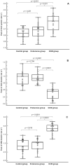
A better understanding of the left ventricle (LV) and right ventricle (RV) functioning would help with the differentiation between athlete's heart and dilated cardiomyopathy (DCM). We aimed to analyse deformation parameters in endurance athletes relative to patients with DCM using cardiac magnetic resonance feature tracking (CMR-FT). The study included males of a similar age: 22 ultramarathon runners, 22 patients with DCM and 21 sedentary healthy controls (41 ± 9 years). The analysed parameters were peak LV global longitudinal, circumferential and radial strains (GLS, GCS and GRS, respectively); peak LV torsion; peak RV GLS. The peak LV GLS was similar in controls and athletes, but lower in DCM (p < 0.0001). Peak LV GCS and GRS decreased from controls to DCM (both p < 0.0001). The best value for differentiation between DCM and other groups was found for the LV ejection fraction (area under the curve (AUC) = 0.990, p = 0.0001, with 90.9% sensitivity and 100% specificity for ≤53%) and the peak LV GRS diastolic rate (AUC = 0.987, p = 0.0001, with 100% sensitivity and 88.4% specificity for >-1.27 s-1). The peak LV GRS diastolic rate was the only independent predictor of DCM (p = 0.003). Distinctive deformation patterns that were typical for each of the analysed groups existed and can help to differentiate between athlete's heart, a nonathletic heart and a dilated cardiomyopathy.
Keywords: athlete’s heart; cardiac magnetic resonance; dilated cardiomyopathy; feature tracking.
Conflict of interest statement
The authors declare no conflict of interest.
Figures


Similar articles
-
Cardiac Magnetic Resonance Feature-Tracking in Olympic Athletes: a myocardial deformation analysis.Eur J Prev Cardiol. 2025 Feb 6:zwaf042. doi: 10.1093/eurjpc/zwaf042. Online ahead of print. Eur J Prev Cardiol. 2025. PMID: 39910863
-
Myocardial mechanics in dilated cardiomyopathy: prognostic value of left ventricular torsion and strain.J Cardiovasc Magn Reson. 2021 Dec 2;23(1):136. doi: 10.1186/s12968-021-00829-x. J Cardiovasc Magn Reson. 2021. PMID: 34852822 Free PMC article.
-
Advanced myocardial characterization in hypertrophic cardiomyopathy: feasibility of CMR-based feature tracking strain analysis in a case-control study.Eur Radiol. 2020 Nov;30(11):6118-6128. doi: 10.1007/s00330-020-06922-6. Epub 2020 Jun 25. Eur Radiol. 2020. PMID: 32588208
-
The Role of Cardiovascular Imaging in the Diagnosis of Athlete's Heart: Navigating the Shades of Grey.J Imaging. 2024 Sep 14;10(9):230. doi: 10.3390/jimaging10090230. J Imaging. 2024. PMID: 39330450 Free PMC article. Review.
-
Value of cardiac magnetic resonance feature-tracking in Arrhythmogenic Cardiomyopathy (ACM): A systematic review and meta-analysis.Int J Cardiol Heart Vasc. 2024 Jul 5;53:101455. doi: 10.1016/j.ijcha.2024.101455. eCollection 2024 Aug. Int J Cardiol Heart Vasc. 2024. PMID: 39228971 Free PMC article. Review.
Cited by
-
Altered Circulating MicroRNA Profiles After Endurance Training: A Cohort Study of Ultramarathon Runners.Front Physiol. 2022 Jan 25;12:792931. doi: 10.3389/fphys.2021.792931. eCollection 2021. Front Physiol. 2022. PMID: 35145424 Free PMC article.
-
Alterations in Circulating MicroRNAs and the Relation of MicroRNAs to Maximal Oxygen Consumption and Intima-Media Thickness in Ultra-Marathon Runners.Int J Environ Res Public Health. 2021 Jul 6;18(14):7234. doi: 10.3390/ijerph18147234. Int J Environ Res Public Health. 2021. PMID: 34299680 Free PMC article.
-
Certainties and Uncertainties of Cardiac Magnetic Resonance Imaging in Athletes.J Cardiovasc Dev Dis. 2022 Oct 20;9(10):361. doi: 10.3390/jcdd9100361. J Cardiovasc Dev Dis. 2022. PMID: 36286312 Free PMC article. Review.
-
Identification of BMP10 as a Novel Gene Contributing to Dilated Cardiomyopathy.Diagnostics (Basel). 2023 Jan 9;13(2):242. doi: 10.3390/diagnostics13020242. Diagnostics (Basel). 2023. PMID: 36673052 Free PMC article.
-
A concise guide of contemporary cardiovascular imaging practices to differentiate athlete's heart in the gray zone.Heart Fail Rev. 2025 Jun 27. doi: 10.1007/s10741-025-10541-y. Online ahead of print. Heart Fail Rev. 2025. PMID: 40576890 Review.
References
-
- Małek Ł.A., Barczuk-Falęcka M., Werys K., Czajkowska A., Mróz A., Witek K., Burrage M., Bakalarski W., Nowicki D., Roik D., et al. Cardiovascular magnetic resonance with parametric mapping in long-term ultra-marathon runners. Eur. J. Radiol. 2019;117:89–94. doi: 10.1016/j.ejrad.2019.06.001. - DOI - PubMed
LinkOut - more resources
Full Text Sources
Other Literature Sources
Miscellaneous

