. 2021 Feb 15;13(2):622.
doi: 10.3390/nu13020622.

Omega-3 Polyunsaturated Fatty Acids Prevent Nonalcoholic Steatohepatitis (NASH) and Stimulate Adipogenesis

Affiliations

Omega-3 Polyunsaturated Fatty Acids Prevent Nonalcoholic Steatohepatitis (NASH) and Stimulate Adipogenesis

Vitor Jacó Antraco et al. Nutrients. .

Abstract

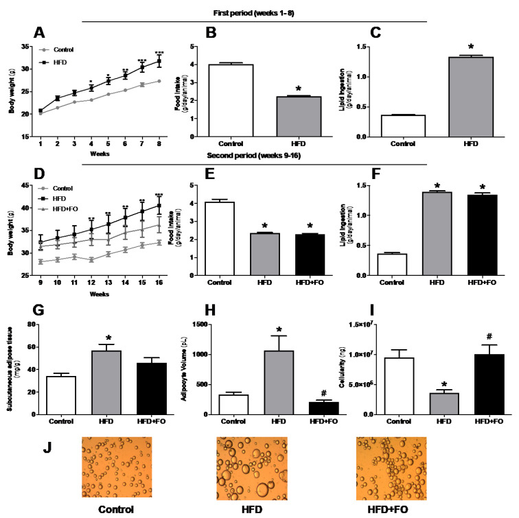
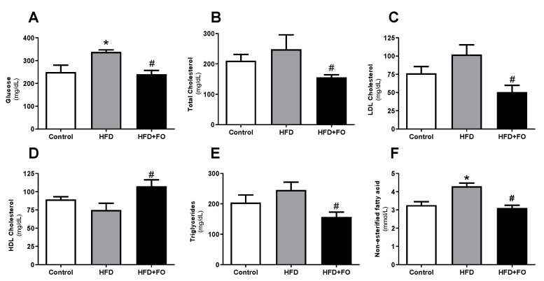
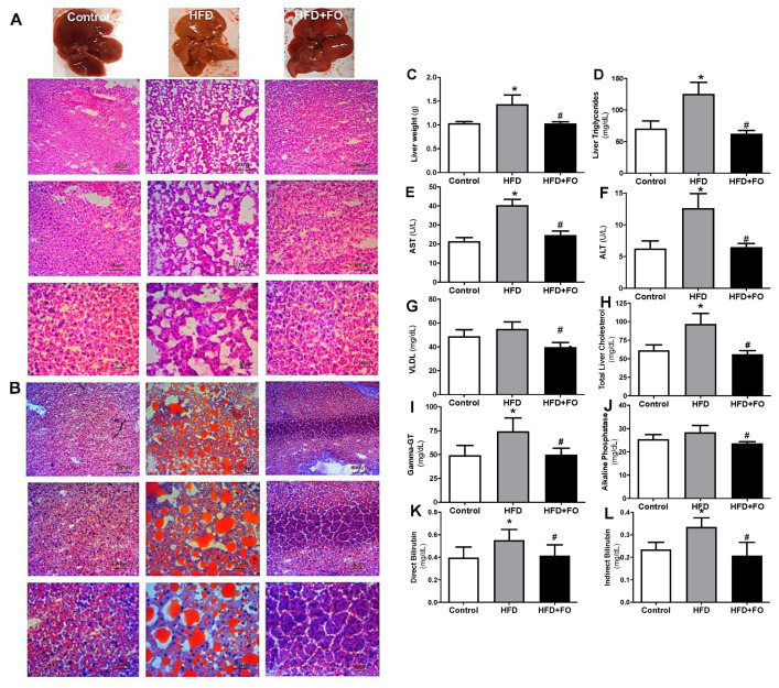
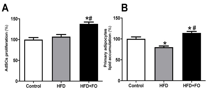
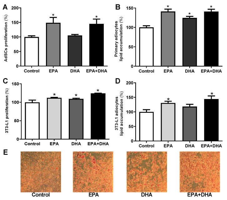
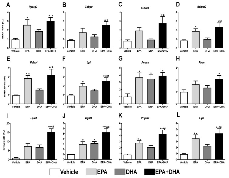
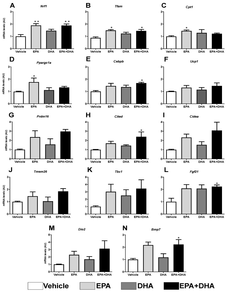
The increasing impact of obesity on global human health intensifies the importance of studies focusing on agents interfering with the metabolism and remodeling not only of the white adipose tissue (WAT) but also of the liver. In the present study, we have addressed the impact of n-3 PUFA in adipose cells' proliferation and adipogenesis, as well as in the hepatic lipid profile and morphology. Mice were induced to obesity by the consumption of a high-fat diet (HFD) for 16 weeks. At the 9th week, the treatment with fish oil (FO) was initiated and maintained until the end of the period. The FO treatment reduced the animals' body mass, plasma lipids, glucose, plasma transaminases, liver mass, triacylglycerol, and cholesterol liver content when compared to animals consuming only HFD. FO also decreased the inguinal (ing) WAT mass, reduced adipocyte volume, increased adipose cellularity (hyperplasia), and increased the proliferation of adipose-derived stromal cells (AdSCs) which corroborates the increment in the proliferation of 3T3-L1 pre-adipocytes or AdSCs treated in vitro with n-3 PUFA. After submitting the in vitro treated (n-3 PUFA) cells, 3T3-L1 and AdSCs, to an adipogenic cocktail, there was an increase in the mRNA expression of adipogenic transcriptional factors and other late adipocyte markers, as well as an increase in lipid accumulation when compared to not treated cells. Finally, the expression of browning-related genes was also higher in the n-3 PUFA treated group. We conclude that n-3 PUFA exerts an attenuating effect on body mass, dyslipidemia, and hepatic steatosis induced by HFD. FO treatment led to decreasing adiposity and adipocyte hypertrophy in ingWAT while increasing hyperplasia. Data suggest that FO treatment might induce recruitment (by increased proliferation and differentiation) of new adipocytes (white and/or beige) to the ingWAT, which is fundamental for the healthy expansion of WAT.

Keywords: AdScs; beige adipocytes; fish oil; liver; obesity.


Conflict of interest statement

The authors declare no conflict of interest. The funders had no role in the design of the study; in the collection, analyses, or interpretation of data; in the writing of the manuscript, or in the decision to publish the results.

Figures

Figure 1
Figure 1
Effects of high-fat diet (HFD) and fish oil (FO) treatment on body weight, food intake, and adiposity. In the first period, mice were fed with a control diet (control group) or HFD. In the second period, the diets were maintained and water (CO and HFD groups) or fish oil (HFD + FO group) were delivered by orogastric gavage. (A,D) body weight (g); (B,E) food intake (g/day/animal); (C,F) lipid ingestion (g/day/animal); (G) subcutaneous adipose tissue (mg/g), (H) ingWAT adipocyte volume (pL); (I) ING cellularity (ng); (J) isolated ING adipocytes photographed under optic microscope (×100 magnification). Results were analyzed by one-way ANOVA and Tukey post-test. Values are mean ± SEM (n = 10–20). * p < 0.05.
Figure 2
Figure 2
Plasma biochemical profile: (A) fasting glucose (mg/dL), (B) total cholesterol (mg/dL), (C) LDL cholesterol (mg/dL), (D) HDL cholesterol (mg/dL), (E) triglycerides (mg/dL), and (F) NEFA (mmol/L), in animals with diet-induced obesity for 16 weeks. The animals received control diet (Control group) or hyperlipidic diet without (HFD group) or with fish oil supplementation in the last eight weeks (HFD + FO group). Values expressed as mean ± SEM (n = 10–20). * p < 0.05 vs. Control; # p < 0.01 vs. HFD; one-way ANOVA and Tukey post-test.
Figure 3
Figure 3
Macroscopic and histological comparison after (A) H&E and (B) oil red O staining for visualization of hepatic lipid accumulation; (C) absolute weight of the liver (g); (D) liver triglycerides (mg/dL); (E) plasma AST (U/L); (F) plasma ALT (U/L); (G) VLDL-c (mg/dL); (H) liver total cholesterol (mg/dL); (I), plasma Gamma-GT (mg/dL); (J) plasma alkaline phosphatase (mg/dL); (K) direct bilirubin (mg/dL); (L) indirect bilirubin (mg/dL). Mice received control diet (Control) or hyperlipidic diet without (HFD) or with fish oil treatment (HFD + FO). Values expressed as mean ± SEM (n = 10–20). * p < 0.05 vs. Control, # p < 0.05 vs. HFD; one-way ANOVA and Tukey post-test.
Figure 4
Figure 4
AdSCs proliferation and adipocyte differentiation. (A) the proliferation and (B) evaluation of intracellular lipid accumulation determined after oil red O staining and estimated by spectrophotometry after induction of adipogenesis of primary pre-adipocytes from control diet (Control) or hyperlipid diet (HFD) mice treated with fish oil (HFD + FO). The pre-adipocytes were obtained from the cell fraction of the vascular stroma of the WAT inguinal. Values expressed as mean ± SEM (n = 6–15). * p < 0.001 vs. Control, # p < 0.01 vs. HFD; one-way ANOVA and Tukey post-test.
Figure 5
Figure 5
Proliferation, evaluation of cell lipid accumulation determined by oil red O staining after induction of adipogenesis in primary mice pre-adipocytes (A,B) and 3T3-L1 (CE) cultivated in vitro in the presence or not (control) of 50 µM of eicosapentaenoic acid (EPA, C20:5 n-3), docosahexaenoic (DHA, C22:6 n-3), its association (EPA/DHA) in a 5:1 ratio (42 µM EPA: 8 µM DHA). Values expressed as mean ± SEM (n = 6–15). * p < 0.05 vs. Control; one-way ANOVA and Tukey post-test.
Figure 6
Figure 6
Evaluation of gene expression 8 days after differentiation of 3T3-L1 preadipocytes by RT-PCR in the presence or not (control) of 50 µM of eicosapentaenoic acids (EPA, C20:5 n-3), docosaexaenoic (DHA, C22:6 n-3) or their association in a 5:1 ratio (42 µM EPA: 8 µM DHA). (A) Ppar-gamma; (B) C/ebp-alpha; (C) Sic2a4 (Glut-4); (D) AdipoQ (Adiponectin); (E) Fabp4; (F) Lpl; (G) Acaca (Acc1); (H) Fasn (FAS); (I) Lpin1 (Lipin); (J) Dgat1; (K) Pnpl2 (Atgl); (L) Lipe (Hsl). Values of mRNA were expressed in relation to the control and corrected by the expression of the constitutive gene 36B4. Values are expressed as mean ± SEM (n = 4–7). * p < 0.05 vs. Control, ** p < 0.01 vs. Control, *** p < 0.001 vs. Control, # p < 0.01 vs. DHA. One-way ANOVA and Tukey post-test.
Figure 7
Figure 7
Evaluation of gene expression of beige adipocyte markers after browning induction six days after differentiation into 3T3-L1 preadipocytes by RT-PCR in the presence or not (control) of 50 µM of eicosapentaenoic acid (EPA, C20:5 n-3), docosaexaenoic (DHA, C22:6 n-3) or their association in a 5:1 ratio (42 µM EPA: 8 µM DHA). (A) Nrf1; (B) Tfam; (C) Cpt1; (D) Ppargc1a (Pgc1-α); (E) Cebpβ; (F) Ucp1; (G) Prdm16; (H) Cited; (I) Cidea; (J) Tmem26; (K) Tbx; (L) Fgf21; (M) Dio2; (N) Bmp7. Values of mRNA were expressed in relation to the control and corrected by the expression of the constitutive gene 36B4. Values expressed as mean ± SEM (n = 4–7). * p < 0.05 vs. Control, ** p < 0.01 vs. Control. One-way ANOVA and Tukey post-test.

References

    1. Chechi K., Nedergaard J., Richard D. Brown adipose tissue as an anti-obesity tissue in humans. Obes. Rev. 2014;15:92–106. doi: 10.1111/obr.12116. - DOI - PubMed
    1. González-Muniesa P., Mártinez-González M.A., Hu F.B., Després J.P., Matsuzawa Y., Loos R.J.F., Moreno L.A., Bray G.A., Martinez J.A. Obesity. Nat. Rev. Dis. Prim. 2017;3:1–18. doi: 10.1038/nrdp.2017.34. - DOI - PubMed
    1. Albuquerque D., Stice E., Rodríguez-López R., Manco L., Nóbrega C. Current review of genetics of human obesity: From molecular mechanisms to an evolutionary perspective. Mol. Genet. Genom. 2015;290:1191–1221. doi: 10.1007/s00438-015-1015-9. - DOI - PubMed
    1. Than N.N., Newsome P.N. A concise review of non-alcoholic fatty liver disease. Atherosclerosis. 2015;239:192–202. doi: 10.1016/j.atherosclerosis.2015.01.001. - DOI - PubMed
    1. Xiang Z., Chen Y., Ma K., Ye Y., Zheng L., Li Y., Jin X. The role of ursodeoxycholic acid in non-alcoholic steatohepatitis: A systematic review. BMC Gastroenterol. 2013;13:140. doi: 10.1186/1471-230X-13-140. - DOI - PMC - PubMed

LinkOut - more resources