. 2021 Feb 15;10(2):398.
doi: 10.3390/cells10020398.

Sensitivity of the Natriuretic Peptide/cGMP System to Hyperammonaemia in Rat C6 Glioma Cells and GPNT Brain Endothelial Cells

Affiliations

Sensitivity of the Natriuretic Peptide/cGMP System to Hyperammonaemia in Rat C6 Glioma Cells and GPNT Brain Endothelial Cells

Jacob T Regan et al. Cells. .

Abstract

C-type natriuretic peptide (CNP) is the major natriuretic peptide of the central nervous system and acts via its selective guanylyl cyclase-B (GC-B) receptor to regulate cGMP production in neurons, astrocytes and endothelial cells. CNP is implicated in the regulation of neurogenesis, axonal bifurcation, as well as learning and memory. Several neurological disorders result in toxic concentrations of ammonia (hyperammonaemia), which can adversely affect astrocyte function. However, the relationship between CNP and hyperammonaemia is poorly understood. Here, we examine the molecular and pharmacological control of CNP in rat C6 glioma cells and rat GPNT brain endothelial cells, under conditions of hyperammonaemia. Concentration-dependent inhibition of C6 glioma cell proliferation by hyperammonaemia was unaffected by CNP co-treatment. Furthermore, hyperammonaemia pre-treatment (for 1 h and 24 h) caused a significant inhibition in subsequent CNP-stimulated cGMP accumulation in both C6 and GPNT cells, whereas nitric-oxide-dependent cGMP accumulation was not affected. CNP-stimulated cGMP efflux from C6 glioma cells was significantly reduced under conditions of hyperammonaemia, potentially via a mechanism involving changed in phosphodiesterase expression. Hyperammonaemia-stimulated ROS production was unaffected by CNP but enhanced by a nitric oxide donor in C6 cells. Extracellular vesicle production from C6 cells was enhanced by hyperammonaemia, and these vesicles caused impaired CNP-stimulated cGMP signalling in GPNT cells. Collectively, these data demonstrate functional interaction between CNP signalling and hyperammonaemia in C6 glioma and GPNT cells, but the exact mechanisms remain to be established.

Keywords: astrocyte; cGMP; endothelial cells; extracellular vesicles; hyperammonaemia; natriuretic peptides; neuroendocrinology.


Conflict of interest statement

The authors declare no conflict of interest.

Figures

Figure 1
Figure 1
Molecular and pharmacological characterization of natriuretic peptide system in rat C6 glioma cells. (A) Multiplex RT-qPCR was performed on RNA extracted from rat brain tissue or C6 cells. The data shown are mean relative gene expression, normalized to Actb, of 3 to 6 individual RNA extractions. (B) Total cGMP accumulation in C6 cells treated with 0 or 100 nM ANP or CNP for 1 h in physiological saline solution containing 1 mM IBMX, before measuring with a commercially available cGMP-EIA kit (R&D Systems) as described previously. Data shown are means ± SEM pooled from three independent experiments, each performed in triplicate. **** p < 0.0001, significantly different from basal. (C) Concentration-dependent effects of CNP on cGMP accumulation in C6 cells. Cells were treated with the indicated concentrations of CNP in PSS containing 1 mM IBMX for 1 h. Data shown are means ± SEM, representative of three independent experiments, each performed in triplicate. (D) C6 cells were pre-treated in media containing 1% (v/v) FCS and 0.10 mM NH4Cl (HA), or 10 ng/mL of either IL1β, IL6, TNFα or CRP for 24 h prior to subsequent stimulation with 100 nM CNP in the presence of 1 mM IBMX for 15 min. The data shown are means ± SEM of five to twelve individual stimulations, expressed as the percentage of the control response to CNP * p < 0.05, significantly different from control response to CNP. The dotted line indicates basal cGMP concentration. (E) C6 cells were pre-treated in media containing 1% (v/v) FCS and 0, 1, 5 or 10 mM NH4Cl for 24 h prior to subsequent stimulation with 100 nM CNP in the presence of 1 mM IBMX for 15 min. The data shown are means ± SEM of four to ten independent experiments, each performed in triplicate and are expressed as the percentage of the control response to CNP. * p < 0.05, ** p < 0.01, significantly different from control response to CNP.
Figure 2
Figure 2
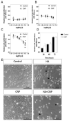
Effects of hyperammonaemia and CNP on cell proliferation and morphology in rat C6 glioma cells. C6 cells were treated with the indicated concentrations of NH4Cl (log mM) (as an NH3 donor) in the absence or presence of 100 nM CNP for (A) 24 h, (B) 48 h or (C) 72 h, before being fixed in paraformaldehyde, and stained for crystal violet assay. The data shown are means ± SEM pooled from 3 independent experiments, each performed with 8 replicates. * p < 0.05, ** p < 0.01, significantly different from untreated cells. (D) Cell viability in C6 cells in the presence of 0 or 100 nM CNP over 72 h. The data shown are means ± SEM pooled from 3 independent experiments, each performed with 8 replicates. (E) Representative microscopy images of C6 cells treated for 72 h with 0 (control) or 10 mM NH4Cl (HA), in the presence of 100 nM CNP alone (CNP) or in combination (HA+CNP). Arrow heads indicated rounded cells.
Figure 3
Figure 3
Effects of hyperammonaemia on CNP-stimulated cGMP accumulation in rat C6 glioma cells. Cells were initially treated in media containing 1% (v/v) FCS and 0 or 10 mM NH4Cl (HA) for either (A) 1 h or (B) 24 h, prior to subsequent stimulation with 100 nM CNP in the presence of 1 mM IBMX for up to 15 min. The data shown are means ± SEM of five to seven independent experiments, each performed in triplicate and are expressed as the percentage of the control; * p < 0.05, ** p < 0.01, significantly different from control response to CNP. (C) Linear accumulation of cGMP between 5 and 15 min of CNP stimulation. Data shown are normalized to cGMP accumulation after 5 min, expressed as means ± SEM of five to seven independent experiments. ** p < 0.01, significantly deviates from zero.
Figure 4
Figure 4
Effects of hyperammonaemia on SNP-stimulated cGMP accumulation in rat C6 glioma cells. Cells were initially treated in media containing 1% (v/v) FCS and 0 or 10 mM NH4Cl (HA) for either (A) 1 h or (B) 24 h, prior to subsequent stimulation with 1 mM SNP in the presence of 1 mM IBMX for up to 15 min. The data shown are means ± SEM of three independent experiments, each performed in triplicate and are expressed as the percentage of the control response to SNP. (C) Linear accumulation of cGMP between 5 and 15 min of SNP stimulation. Data shown are normalized to cGMP accumulation after 5 min, expressed as means ± SEM of three independent experiments. *** p < 0.001, ** p < 0.01, significantly deviates from zero.
Figure 5
Figure 5
Effects of hyperammonaemia on CNP-stimulated cGMP efflux rat C6 glioma cells. C6 cells were treated in media containing 1% (v/v) FCS and either 0 or 10 mM NH4Cl, (HA) in the absence or presence of 100 nM CNP, for up to 72 h. Spent media were collected at the indicated time points and analysed for cGMP content. Data shown are means ± SEM of 6 to 8 independent experiments, each performed in triplicate, and normalized to basal at each time point. **** p < 0.0001, significantly different to control response to CNP.
Figure 6
Figure 6
Effects of hyperammonaemia on cGMP-associated, and astrocyte-enriched gene expression in rat C6 glioma cells. Cells were initially treated in media containing 1% (v/v) FCS and 0 or 10 mM NH4Cl (HA) for 48 h, prior to extracting total RNA. Multiplex RT-qPCR assays were performed on RNA extracted from C6 cells to detect (A) transcripts encoding phosphodiesterase enzymes, (B) other cGMP-associated genes and astrocyte-enriched transcripts. The data shown are the medians (with 5 to 95% confidence intervals) of relative gene expression, normalized to Actb and expressed as fold change over basal, from 4 to 12 individual RNA extractions. * p < 0.05, ** p < 0.01, *** p < 0.001, **** p < 0.0001, significantly different from control.
Figure 7
Figure 7
(AD). Effects of hyperammonaemia on ROS production in rat C6 glioma cells. Cells were pre-treated in media containing 1% (v/v) FCS and 100 µM DHR for 30 min prior to stimulation with either 0, 10 mM NH4Cl (HA), 100 nM CNP, 1 mM SNP, or combinations of HA+CNP and HA+SNP. ROS production was measured every 30 min for 6 h on a plate-reading spectrophotometer at 500 nm, with a final reading taken after 24 h. The data shown are means ± SEM, pooled from 4 to 6 independent experiments, each performed with 16 replicates (n = 4 to n = 6), and normalised to the 24 h control reading. **** p < 0.0001, significantly different from control.
Figure 8
Figure 8
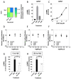
Effects of hyperammonaemia on the natriuretic peptide system in rat GPNT brain endothelial cells. (A) Multiplex RT-qPCR was performed on RNA extracted from rat brain tissue or GPNT cells. The data shown are mean relative gene expression, normalized to Actb, of 2 individual RNA extractions. (B) Total cGMP accumulation in GPNT cells treated with 0 or 100 nM ANP, CNP or 1 mM SNP for 1 h in physiological saline solution containing 1 mM IBMX, before assay. Data shown are means ± SEM of triplicate treatments. (C) Concentration-dependent effects of CNP on cGMP accumulation in C6 cells. Cells were treated with the indicated concentrations of CNP in PSS containing 1 mM IBMX for 1 h. Data shown are means ± SEM pooled from three independent experiments, each performed in triplicate. (DF) Effects of hyperammonaemia and CNP on cell proliferation in GPNT cells. GPNT cells were treated with the indicated concentrations of NH4Cl in the absence or presence of 100 nM CNP or 1 nM SNP for (D) 24 h, (E) 48 h or (F) 72 h, before being fixed in 4% (w/v) paraformaldehyde and stained for crystal violet assay. The data shown are means ± SEM pooled from 3 independent experiments, each performed with 8 replicates. (G,H) Effects of hyperammonaemia on CNP-stimulated cGMP accumulation in GPNT cells. Cells were initially treated in media containing 1% (v/v) FCS and 0 or 10 mM NH4Cl (HA) for either (G) 1 h or (H) 24 h, prior to subsequent stimulation with 100 nM CNP in the presence of 1 mM IBMX for 1 h. The data shown are means ± SEM of five to seven independent stimulations, expressed as the percentage of the control response to CNP; ** p < 0.01, *** p < 0.001, **** p < 0.0001, significantly different from untreated (control), or from the control CNP response to CNP (brackets).
Figure 9
Figure 9
Effects of hyperammonaemia on extracellular vesicle production from C6 cells, and their functional effects on GPNT cells. (A) Extracellular vesicles were harvested from spent media from C6 cells treated with 0 or 10 mM NH4Cl for up to 72 h. These vesicles were stained with Cy7-labelled Annexin-5 and analysed by FACS. Data shown are representative FACS dot plots, showing gating parameters. (B,C) Comparison of Annexin-5-positive extracellular vesicles, and total extracellular vesicles from C6 cells treated with 0 or 10 mM NH4Cl. Data shown are medians (with 5 to 95% confidence intervals) of events, normalized to % of control treated cells, from 3 to 4 independent experiments. * p < 0.05, significantly different from control). (D) Effects of C6-derived extracellular vesicles on CNP-stimulated cGMP accumulation in GPNT cells. GPNT cells were treated with extracellular vesicles from control or NH4Cl-treated C6 cells, for 24 h, prior to subsequent stimulation with 100 nM CNP in the presence of 1 mM IBMX for 1 h. The data shown are means ± SEM from 6 to 9 independent stimulations. **** p < 0.0001, ** p < 0.01 significantly different from unstimulated; * p < 0.05, significantly different from CNP response in C6 EV (0 mM HA) cells.

Similar articles

Cited by

References

    1. Potter L.R., Abbey-Hosch S., Dickey D.M. Natriuretic peptides, their receptors, and cyclic guanosine monophosphate-dependent signaling functions. Endocr. Rev. 2005;27:47–72. doi: 10.1210/er.2005-0014. - DOI - PubMed
    1. Kuhn M. Molecular physiology of membrane guanylyl cyclase receptors. Physiol. Rev. 2016;96:751–804. doi: 10.1152/physrev.00022.2015. - DOI - PubMed
    1. Fowkes R.C., McArdle C.A. C-type natriuretic peptide: An important neuroendocrine regulator? Trends Endocrinol. Metab. 2000;11:333–338. doi: 10.1016/S1043-2760(00)00288-5. - DOI - PubMed
    1. Rose R.A., Giles W.R. Natriuretic peptide C receptor signalling in the heart and vasculature. J. Physiol. 2008;586:353–366. doi: 10.1113/jphysiol.2007.144253. - DOI - PMC - PubMed
    1. Moyes A.J., Khambata R.S., Villar I., Bubb K.J., Baliga R.S., Lumsden N.G., Xiao F., Gane P.J., Rebstock A.-S., Worthington R.J., et al. Endothelial C-type natriuretic peptide maintains vascular homeostasis. J. Clin. Investig. 2014;124:4039–4051. doi: 10.1172/JCI74281. - DOI - PMC - PubMed

Publication types

LinkOut - more resources