Review
. 2021 Feb 15;11(2):316.
doi: 10.3390/diagnostics11020316.

Role of Endoscopic Ultrasound in the Diagnosis of Pancreatic Neuroendocrine Neoplasms

Affiliations
Review

Role of Endoscopic Ultrasound in the Diagnosis of Pancreatic Neuroendocrine Neoplasms

Tatsuya Ishii et al. Diagnostics (Basel). .

Abstract

Although pancreatic neuroendocrine neoplasms (PNENs) are relatively rare tumors, their number is increasing with advances in diagnostic imaging modalities. Even small lesions that are difficult to detect using computed tomography or magnetic resonance imaging can now be detected with endoscopic ultrasound (EUS). Contrast-enhanced EUS is useful, and not only diagnosis but also malignancy detection has become possible by evaluating the vascularity of tumors. Pathological diagnosis using EUS with fine-needle aspiration (EUS-FNA) is useful when diagnostic imaging is difficult. EUS-FNA can also be used to evaluate the grade of malignancy. Pooling the data of the studies that compared the PNENs grading between EUS-FNA samples and surgical specimens showed a concordance rate of 77.5% (κ-statistic = 0.65, 95% confidence interval = 0.59-0.71, p < 0.01). Stratified analysis for small tumor size (2 cm) showed that the concordance rate was 84.5% and the kappa correlation index was 0.59 (95% confidence interval = 0.43-0.74, p < 0.01). The evolution of ultrasound imaging technologies such as contrast-enhanced and elastography and the artificial intelligence that analyzes them, the evolution of needles, and genetic analysis, will further develop the diagnosis and treatment of PNENs in the future.

Keywords: endoscopic ultrasound; pancreatic neuroendocrine neoplasms; pancreatic tumor.


Conflict of interest statement

A.K. received honoraria as a lecture fee from Olympus Co., Tokyo, Japan. The sponsors had no role in the design, execution, interpretation, or writing of the study. The other authors declare no conflicts of interest relevant to this article.

Figures

Figure 1
Figure 1
(a) B-mode endoscopic ultrasound (EUS): a circular hypoechoic mass is seen in the body of the pancreas (yellow arrow). (b) Contrast-enhanced EUS: the mass shows early enhancement compared with the surrounding pancreatic parenchyma (yellow arrow).
Figure 2
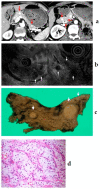
Figure 2
(a) Contrast-enhanced computed tomography (CT): a mass lesion with early hyperenhancement is seen in the tail of the pancreas (red arrow), but there are no other obvious lesions. (b) Diffusion-weighted magnetic resonance imaging (MRI): the mass in the pancreatic tail shows reduced diffusion. (c) Endoscopic ultrasound (EUS): in addition to the mass (☆) revealed by CT/MRI, many small hypoechoic masses are observed (white arrow). (d,e) Resected specimen: the main lesion (☆) is an 11 mm neuroendocrine neoplasm (NEN) G1, but multiple tumors with diameters of 1–3 mm are observed in the surrounding pancreas (white and yellow arrow) (20×). They are multiple NENs associated with multiple endocrine neoplasia type 1 (MEN-1).
Figure 3
Figure 3
(a) Contrast-enhanced computed tomography (CT): a cystic lesion is seen on the pancreatic head. (b) Magnetic resonance imaging: fluid–fluid level formation is shown. (c) B-mode endoscopic ultrasound (EUS): a cystic lesion with fluid–fluid level formation and a thickened wall is seen. (d) Contrast-enhanced EUS: the wall is hyperenhanced compared with the surrounding pancreatic parenchyma. (e,f) Resected specimen: neuroendocrine neoplasm G1 (100×).
Figure 4
Figure 4
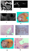
(a) Contrast-enhanced computed tomography (CT): a mass lesion with early hyperenhancement is seen in the pancreatic body (red arrow), but there are no other obvious lesions. (b) Magnetic resonance imaging: the main pancreatic duct (MPD) in the pancreatic body is extensively narrowed, and the caudal duct is dilated. (c) Endoscopic ultrasound (EUS): a circular hypoechoic mass in the pancreatic body. Pancreatic duct stenosis is observed even in the absence of mass. (df) 6-mm neuroendocrine neoplasm G1 (red arrow), serotonin positive, with stromal fibrosis (40×). (g,h) Pancreatic duct stenosis due to stromal fibrosis upstream of the tumor (40×).
Figure 5
Figure 5
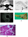
(a) Contrast-enhanced computed tomography (CT): an ill-defined mass (arrow) with a hypovascular enhancement pattern. The dilated pancreatic duct is notable. (b) Endoscopic ultrasound (EUS): irregular margins, unclear boundaries, and heterogeneous hypoechoic masses are shown. (ce) Resected specimen: Ki-67 > 20%, neuroendocrine neoplasm G3 (100×).
Figure 6
Figure 6
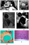
(a) Contrast-enhanced computed tomography (CT): a tumor (arrow) with calcification components is shown at the pancreatic head. (b) Endoscopic ultrasound (EUS): a well-defined mass with a heterogeneous appearance and peripheral rim calcification with posterior acoustic shadowing. (c,d) The imaging findings suggest a solid pseudopapillary neoplasm, but pathologically, it is a neuroendocrine tumor G1 (100×).
Figure 7
Figure 7
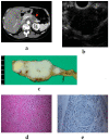
(a) Contrast-enhanced computed tomography (CT): many lesions with early hyperenhancement are seen in the head, body, and tail of the pancreas (red arrow). (b) Endoscopic ultrasound (EUS): many small hypoechoic masses are observed (white arrow). (c,d) Resected specimen: multiple tumors are observed throughout the pancreas (white arrow); they were clear cell carcinomas (400×). The patient had undergone left radical nephrectomy for renal cell carcinoma eight years ago.

Similar articles

Cited by

References

    1. Rösch T., Lightdale C.J., Botet J.F., Boyce G.A., Sivak M.V., Jr., Yasuda K., Heyder N., Palazzo L., Dancygier H., Schusdziarra V. Localization of pancreatic endocrine tumors by endoscopic ultrasonography. N. Engl. J. Med. 1992;326:1721–1726. doi: 10.1056/NEJM199206253262601. - DOI - PubMed
    1. Katanuma A., Maguchi H., Osanai M., Takahashi K. The difference in the capability of delineation between convex and radial arrayed echoendoscope for pancreas and biliary tract; case reports from the standpoint of both convex and radial arrayed echoendoscope. Dig. Endosc. 2011;23:2–8. doi: 10.1111/j.1443-1661.2011.01131.x. - DOI - PubMed
    1. Kaneko M., Katanuma A., Maguchi H., Takahashi K., Osanai M., Yane K., Hashigo S., Harada R., Kato S., Kato R. Prospective, randomized, comparative study of delineation capability of radial scanning and curved linear array endoscopic ultrasound for the pancreaticobiliary region. Endosc. Int. Open. 2014;2:E160. doi: 10.1055/s-0034-1377384. - DOI - PMC - PubMed
    1. Puli S.R., Kalva N., Bechtold M.L., Pamulaparthy S.R., Cashman M.D., Estes N.C., Pearl R.H., Volmar F.-H., Dillon S., Shekleton M.F. Diagnostic accuracy of endoscopic ultrasound in pancreatic neuroendocrine tumors: A systematic review and meta analysis. World J. Gastroenterol. WJG. 2013;19:3678. doi: 10.3748/wjg.v19.i23.3678. - DOI - PMC - PubMed
    1. Manta R., Nardi E., Pagano N., Ricci C., Sica M., Castellani D., Bertani H., Piccoli M., Mullineris B., Tringali A. Pre-operative diagnosis of pancreatic neuroendocrine tumors with endoscopic ultrasonography and computed tomography in a large series. J. Gastrointestin. Liver Dis. 2016;25:317–321. doi: 10.15403/jgld.2014.1121.253.ned. - DOI - PubMed

LinkOut - more resources