Review
. 2021 Feb 23;10(2):167.
doi: 10.3390/biology10020167.

Proteolytic Cleavages in the VEGF Family: Generating Diversity among Angiogenic VEGFs, Essential for the Activation of Lymphangiogenic VEGFs

Affiliations
Review

Proteolytic Cleavages in the VEGF Family: Generating Diversity among Angiogenic VEGFs, Essential for the Activation of Lymphangiogenic VEGFs

Jaana Künnapuu et al. Biology (Basel). .

Abstract

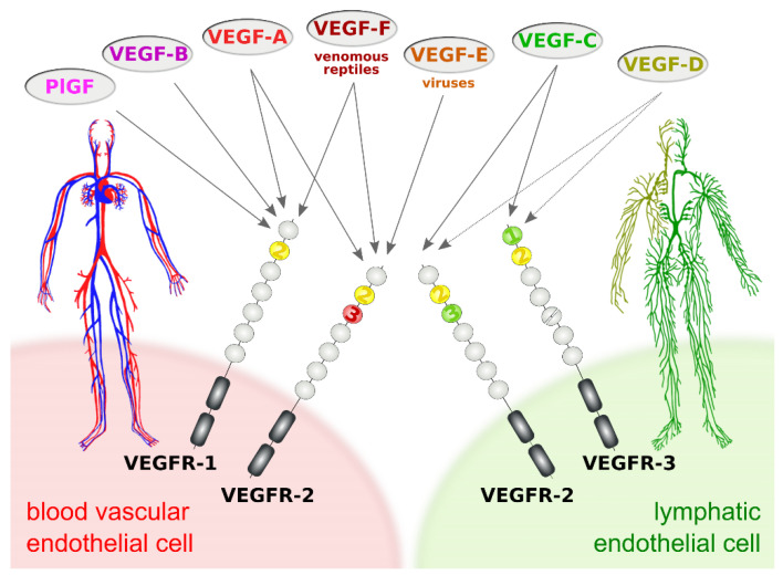
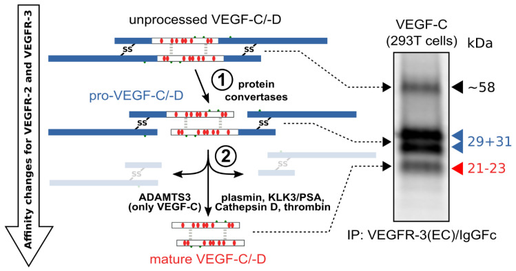
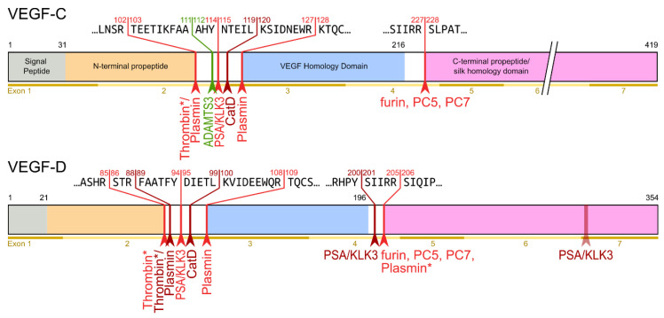
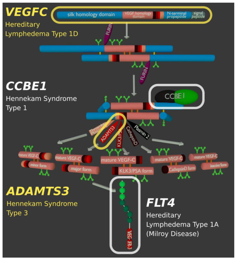
Specific proteolytic cleavages turn on, modify, or turn off the activity of vascular endothelial growth factors (VEGFs). Proteolysis is most prominent among the lymph-angiogenic VEGF-C and VEGF-D, which are synthesized as precursors that need to undergo enzymatic removal of their C- and N-terminal propeptides before they can activate their receptors. At least five different proteases mediate the activating cleavage of VEGF-C: plasmin, ADAMTS3, prostate-specific antigen, cathepsin D, and thrombin. All of these proteases except for ADAMTS3 can also activate VEGF-D. Processing by different proteases results in distinct forms of the "mature" growth factors, which differ in affinity and receptor activation potential. The "default" VEGF-C-activating enzyme ADAMTS3 does not activate VEGF-D, and therefore, VEGF-C and VEGF-D do function in different contexts. VEGF-C itself is also regulated in different contexts by distinct proteases. During embryonic development, ADAMTS3 activates VEGF-C. The other activating proteases are likely important for non-developmental lymphangiogenesis during, e.g., tissue regeneration, inflammation, immune response, and pathological tumor-associated lymphangiogenesis. The better we understand these events at the molecular level, the greater our chances of developing successful therapies targeting VEGF-C and VEGF-D for diseases involving the lymphatics such as lymphedema or cancer.

Keywords: ADAMTS3; CCBE1; KLK3; PlGF; VEGF-A; VEGF-B; VEGF-C; VEGF-D; angiogenesis; cathepsin D; lymphangiogenesis; lymphedema; metastasis; plasmin; prostate-specific antigen (PSA); proteases; proteolytic activation; thrombin; vascular biology; vascular endothelial growth factors (VEGFs); wound healing.


Conflict of interest statement

The authors declare no conflict of interest.

Figures

Figure 1
Figure 1
Vascular endothelial growth factors (VEGFs) act on blood vessels and/or lymphatic vessels depending on their affinities towards VEGF receptors 1, -2, and -3. VEGFR-2 is expressed on both blood and lymphatic endothelium. In principle, growth factors that do activate VEGFR-2 can promote both the growth of blood vessels (angiogenesis) and lymphatic vessels (lymphangiogenesis). VEGF-E and VEGF-F are not of human origin: VEGF-E genes are found in viral genomes, and VEGF-F is a snake venom component. All receptor-growth factor interactions require the extracellular domain 2 of the VEGF receptors (shown in yellow) [6,7,8,9]. Domain 3 of VEGFR-2 is important for the interaction of VEGFR-2 with both VEGF-A [7] and VEGF-C [8], and domain 1 of VEGFR-3 is important for the interaction of VEGF-C with VEGFR-3 [9].
Figure 2
Figure 2
Most diversity among the hemangiogenic VEGFs is achieved by alternative splicing. Nevertheless, proteolytic processing of VEGF-A (A) [19] and placenta growth factor (PlGF) [32] (C) can convert the longer, heparin-binding isoforms into more soluble shorter species. (B) VEGF-B is a special case. Alternative splicing results in two isoforms that translate the same nucleotide sequence in two different frames resulting in a heparin-binding and a soluble isoform [33,34]. Due to the near-perfect cleavage context [35], thrombin has been suspected to be the responsible protease for VEGF-B186 cleavage [36]. Prothrombin is indeed expressed by 293T cells [37], in which the cleavage has been demonstrated [33]. Plasmin cleaves VEGF-B186 at at least four different sites, of which the two most likely predicted sites are indicated. Importantly, the predicted plasmin cleavage between Arg137 and Ala138 removes the interaction epitope for neuropilin-1 binding [33] Semi-transparent, blurry arrows indicate cleavages, for which only the approximate position is known. The figure shows only the most sensitive site from the plasmin cleavages of VEGF-A since prolonged incubation results in progressing degradation [30]. VEGF-B186 appears to be progressively degraded by plasmin as well [33]. For VEGF-A and PlGF, the numbering is according to the longest shown isoform. VEGF-A is cleaved not only by MMP3 but also in a similar fashion by MMP7, MMP9, MMP19, and - less efficiently - by MMP1 and MMP16 [30].
Figure 3
Figure 3
Two proteolytic cleavages are needed to activate VEGF-C and VEGF-D. The first cleavage, by protein convertases, is constitutive and intracellular. The second is highly regulated and happens after secretion of the pro-forms. Many different enzymes have been shown to catalyze the second cleavage, but the primary activating protease of VEGF-C in mammalian developmental lymphangiogenesis is A Disintegrin and Metalloprotease With Thrombospondin Motifs-3 (ADAMTS3). The immunoprecipitation (IP) of transfected 293T cells with a VEGFR-3(EC)/IgGFc fusion protein pulls down the 58 kDa full-length VEGF-C, the pro-VEGF-C peptides of 31 kDa and 29 kDa, and the mature VEGF-C. Proteins were resolved under reducing conditions by SDS-PAGE.
Figure 4
Figure 4
Human VEGF-C and -D are processed in a very similar fashion. The major difference between VEGF-C and -D is that ADAMTS3 activates VEGF-C, but not VEGF-D. This is one of the reasons why ADAMTS3 and VEGF-C are essential for lymphatic development and embryonic survival [14,42], whereas VEGF-D deletion in mice is well tolerated [53]. While the figure shows the exon structure of VEGF-C and -D, mRNA splice isoforms have only been reported for murine Vegfc [54]. The detected splice variants do not contain the full VEGF homology domain and are therefore not shown here. *Cleavage site is only predicted based on the amino acid context.
Figure 5
Figure 5
Biochemically, the group of lymphangiogenic activator enzymes is diverse. It includes a metalloproteinase (ADAMTS3), serine proteases (prostate-specific antigen (PSA)/KLK3, thrombin, and plasmin), and an aspartic protease (cathepsin D). VEGF-C and VEGF-D share all activating enzymes except for the most important one: ADAMTS3. ADAMTS3 is exclusive for VEGF-C and required for the physiologic activation of VEGF-C during developmental lymphangiogenesis [41,42,43]. VEGF-C and VEGF-D are differently affected by proteolytic processing. With progressing processing, VEGF-C largely maintains its lymphangiogenic properties but loses its angiogenic properties quickly. VEGF-D behaves precisely the opposite way: processing with Cathepsin D almost completely abolishes its lymphangiogenic properties and fully unmasks its angiogenic properties [45]. Extensive exposure of both VEGF-C and VEGF-D to plasmin abolishes all VEGFR-2 and VEGFR-3 binding properties.
Figure 6
Figure 6
Genes of the VEGF-C/VEGFR-3 signaling pathway known to be involved in hereditary lymphedema. Although different components of the signaling pathway can be affected, the largest fraction of cases are caused by mutations in the FLT4 gene, which codes for the VEGF-C receptor VEGFR-3.

Similar articles

Cited by

References

    1. de Groof A., Guelen L., Deijs M., van der Wal Y., Miyata M., Ng K.S., van Grinsven L., Simmelink B., Biermann Y., Grisez L., et al. A Novel Virus Causes Scale Drop Disease in Lates Calcarifer. PLOS Pathog. 2015;11:e1005074. doi: 10.1371/journal.ppat.1005074. - DOI - PMC - PubMed
    1. Lyttle D.J., Fraser K.M., Fleming S.B., Mercer A.A., Robinson A.J. Homologs of Vascular Endothelial Growth Factor Are Encoded by the Poxvirus Orf Virus. J. Virol. 1994;68:84–92. doi: 10.1128/JVI.68.1.84-92.1994. - DOI - PMC - PubMed
    1. Meyer M., Clauss M., Lepple-Wienhues A., Waltenberger J., Augustin H.G., Ziche M., Lanz C., Büttner M., Rziha H.-J., Dehio C. A Novel Vascular Endothelial Growth Factor Encoded by Orf Virus, VEGF-E, Mediates Angiogenesis via Signalling through VEGFR-2 (KDR) but Not VEGFR-1 (Flt-1) Receptor Tyrosine Kinases. EMBO J. 1999;18:363–374. doi: 10.1093/emboj/18.2.363. - DOI - PMC - PubMed
    1. Ogawa S., Oku A., Sawano A., Yamaguchi S., Yazaki Y., Shibuya M. A Novel Type of Vascular Endothelial Growth Factor, VEGF-E (NZ-7 VEGF), Preferentially Utilizes KDR/Flk-1 Receptor and Carries a Potent Mitotic Activity without Heparin-Binding Domain. J. Biol. Chem. 1998;273:31273–31282. doi: 10.1074/jbc.273.47.31273. - DOI - PubMed
    1. Yamazaki Y., Matsunaga Y., Tokunaga Y., Obayashi S., Saito M., Morita T. Snake Venom Vascular Endothelial Growth Factors (VEGF-Fs) Exclusively Vary Their Structures and Functions among Species. J. Biol. Chem. 2009;284:9885–9891. doi: 10.1074/jbc.M809071200. - DOI - PMC - PubMed

LinkOut - more resources