. 2021 Feb 23;10(2):331.
doi: 10.3390/antiox10020331.

Oxidatively Modified LDL Suppresses Lymphangiogenesis via CD36 Signaling

Affiliations

Oxidatively Modified LDL Suppresses Lymphangiogenesis via CD36 Signaling

Bhupesh Singla et al. Antioxidants (Basel). .

Abstract

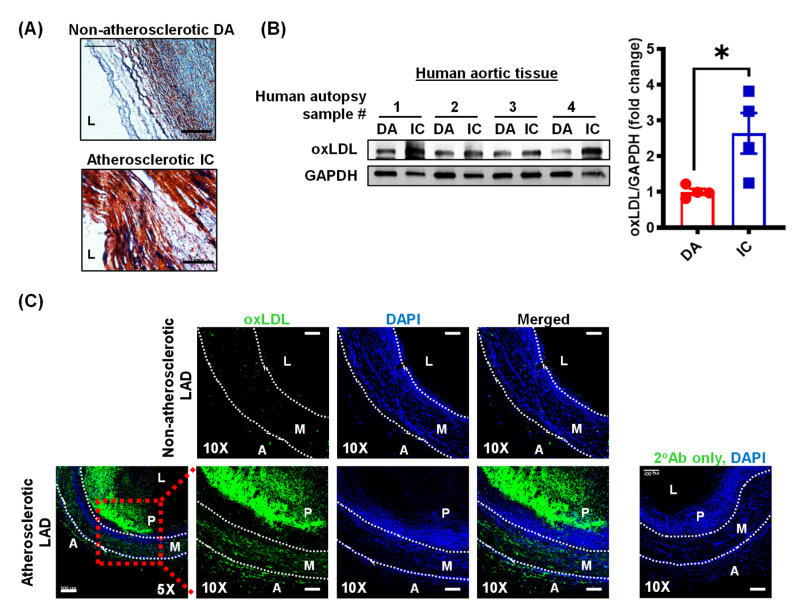
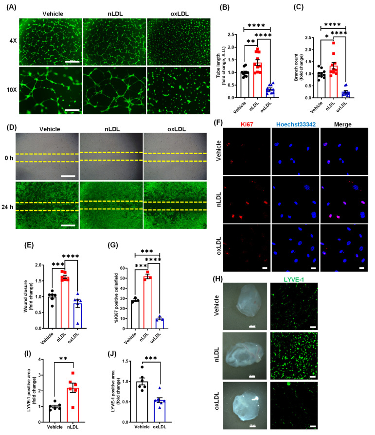
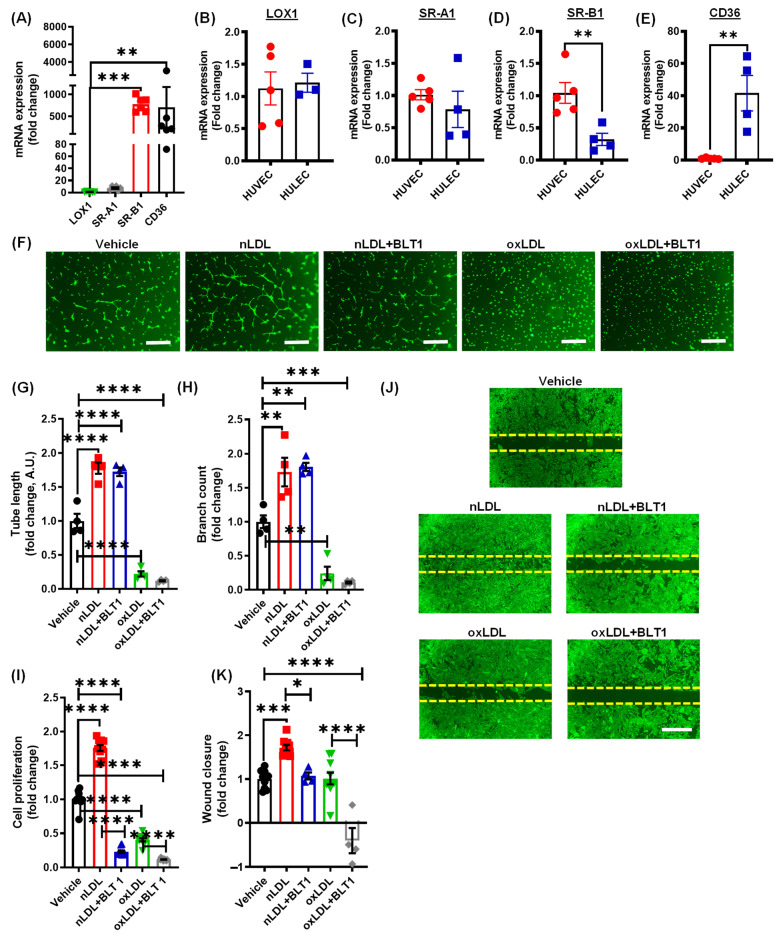
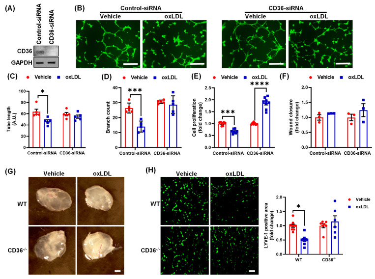
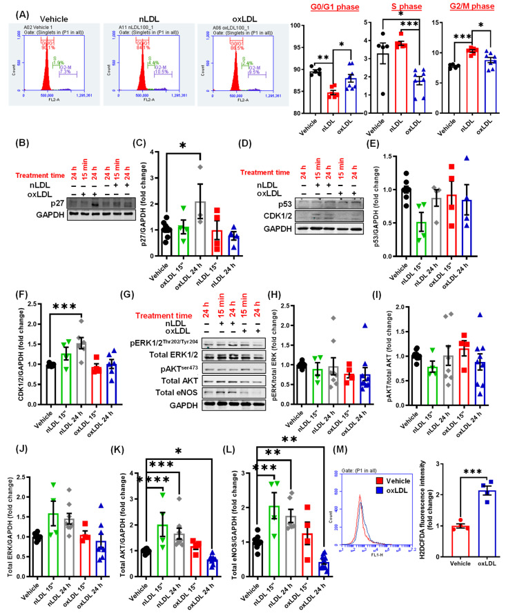
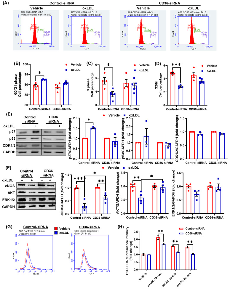
Arterial accumulation of plasma-derived LDL and its subsequent oxidation contributes to atherosclerosis. Lymphatic vessel (LV)-mediated removal of arterial cholesterol has been shown to reduce atherosclerotic lesion formation. However, the precise mechanisms that regulate LV density and function in atherosclerotic vessels remain to be identified. The aim of this study was to investigate the role of native LDL (nLDL) and oxidized LDL (oxLDL) in modulating lymphangiogenesis and underlying molecular mechanisms. Western blotting and immunostaining experiments demonstrated increased oxLDL expression in human atherosclerotic arteries. Furthermore, elevated oxLDL levels were detected in the adventitial layer, where LV are primarily present. Treatment of human lymphatic endothelial cells (LEC) with oxLDL inhibited in vitro tube formation, while nLDL stimulated it. Similar results were observed with Matrigel plug assay in vivo. CD36 deletion in mice and its siRNA-mediated knockdown in LEC prevented oxLDL-induced inhibition of lymphangiogenesis. In addition, oxLDL via CD36 receptor suppressed cell cycle, downregulated AKT and eNOS expression, and increased levels of p27 in LEC. Collectively, these results indicate that oxLDL inhibits lymphangiogenesis via CD36-mediated regulation of AKT/eNOS pathway and cell cycle. These findings suggest that therapeutic blockade of LEC CD36 may promote arterial lymphangiogenesis, leading to increased cholesterol removal from the arterial wall and reduced atherosclerosis.

Keywords: CD36; atherosclerosis; lymphangiogenesis; native LDL; oxidized LDL.


Conflict of interest statement

The authors declare no conflict of interest.

Figures

Figure 1
Figure 1
Oxidized LDL levels are increased in human atherosclerotic arteries. (A) Human aortic tissues (IC and DA) isolated from cadaveric donors were stained with Oil Red O to identify atherosclerotic lesions. Representatives of n = 4 experiments shown. Scale bar: 100 µm. (B) Human aortic tissue lysates were subjected to Western blot analysis for oxLDL and GADPH expression. Representative Western blot images are shown. Bar graph represents mean protein levels along with individual data points calculated using densitometric analysis and expressed as a ratio of oxLDL to GAPDH (n = 4). (C) Atherosclerotic and non-atherosclerotic human LAD coronary arteries were immunostained for oxLDL (green) and nuclei were counterstained with DAPI (blue). Representative images are shown (n = 3 for atherosclerotic LAD and n = 1 for non-atherosclerotic control LAD). Scale bar: 200 µm (leftmost panel). Magnified images of red inset are also shown, scale bar: 100 µm. Data represent the mean ± SEM. * p < 0.05. DA: descending aorta; IC: inner curvature; LAD: left anterior descending; L: lumen; A: adventitia; M: media; and P: plaque.
Figure 2
Figure 2
Oxidized LDL suppresses lymphangiogenesis in vitro and in vivo. (AC) Human LEC in basal media MV2 (0.5% FBS) containing vehicle (PBS), nLDL (100 µg/mL) or oxLDL (100 µg/mL) were seeded in wells of a Matrigel-coated plate and tube formation determined after 6 h. (A) Representative images for LEC tube formation assay are shown. Scale bars: 500 μm for 4× magnification and 200 μm for 10× magnification. Images of random fields were taken, and tube length (B) and the number of branching points (C) quantified (n = 10). All experiments were performed in duplicate. (D,E) LEC migration in response to vehicle, nLDL (100 µg/mL) or oxLDL (100 µg/mL) was investigated after 24 h using Culture-Insert 2 Well 24 (ibidi USA). (D) Representative images of wounds at 0 h and 24 h are shown. Scale bar: 500 μm. (E) Quantification of wound closure (n = 6). (F,G) LEC grown on coverslips were treated with vehicle, nLDL, or oxLDL for 24 h. Cells were fixed and immunostained for Ki67 (red). Nuclei were counterstained with Hoechst 33342 (blue). Images were captured from more than five random fields. (F) Representative images of Ki67 immunostaining. Scale bar: 20 μm. (G) Bar graph represents the mean number of Ki67 positive cells/field (n = 3). (HJ) Wild-type mice were injected s.c. with Matrigel solutions premixed with vehicle, nLDL, or oxLDL. Matrigel plugs were harvested after 2 weeks of implantation. (H) Representative images of harvested Matrigels and LYVE-1 staining of the cross-sections of the Matrigel plugs are shown. Scale bar: 50 μm. (I,J) Quantification of LYVE-1 positive area for H (n = 6). Data represent the mean ± SEM. * p < 0.05; ** p < 0.01; *** p < 0.005; and **** p < 0.001.
Figure 3
Figure 3
Pharmacological blockade of SR-B1 does not rescue oxLDL-induced inhibition of lymphatic endothelial cell proliferation and tube formation. (A) RNA isolated from human dermal LEC was used to determine the relative mRNA expression of LOX-1, SRA1, SRB1 and CD36. GAPDH was used as internal control. Bar graph represents mRNA levels in comparison to LOX1 (gene with the lowest expression) (n = 6). (BE) comparison of mRNA expression of LOX-1 (B), SRA1 (C), SRB1 (D), and CD36 (E) in human LEC (HULEC) and human venous endothelial cells (HUVEC) (n = 4, 5). (FK) LECs were pretreated with vehicle or BLT-1 (10 µM, 1 h), then incubated with vehicle (PBS), nLDL, or oxLDL and tube formation (6 h), cell proliferation (48 h), and cell migration (24 h) investigated as described in Figure 2. (F) Representative images for LEC tube formation assay are shown. Scale bar: 250 μm. Tube length (G) and branching points (H) are quantified (n = 4). (I) Quantification of LEC proliferation using the WST-1 assay (n = 10). (J,K) Representative images of wounds after 24 h and quantification (n = 4–10). Scale bar: 500 μm. Data represent the mean ± SEM. * p < 0.05; ** p < 0.01; *** p < 0.005; and **** p < 0.001.
Figure 4
Figure 4
Oxidized LDL inhibits lymphangiogenesis via CD36. (AF) Human LEC were transfected with control-siRNA or CD36-siRNA. After 48 h, cells were treated with vehicle or oxLDL. (A) Western blot analysis of CD36 protein expression in control and CD36 siRNA-treated LEC. (B) Representative images of LEC tube formation. Scale bar: 200 μm. (C) Quantification of tube length (n = 5). (D) Quantification of branch count (n = 5). (E) Cell proliferation analysis in control and CD36-silenced LEC using the WST-1 assay (n = 7–8). (F) Quantification of cell migration following vehicle and oxLDL treatment (n = 3). (GH) Wild type (WT) and CD36−/− mice were injected subcutaneously (s.c.) with Matrigel solutions premixed with vehicle or oxLDL. (G) Representative images of harvested Matrigel plugs. Scale bar: 50 μm. (H) LYVE-1 immunostaining. Scale bar: 50 μm. Bar graph shows the quantification of LYVE-1 positive area (n = 7–9). Data represent the mean ± SEM. * p < 0.05; *** p < 0.005; and **** p < 0.001.
Figure 5
Figure 5
Oxidized LDL induces cell cycle arrest and inhibits AKT and eNOS expression in LEC. (A) Human LEC were treated with vehicle, nLDL, or oxLDL for 24 h, and cell cycle analysis was performed using flow cytometry. Representative flow cytometry histograms for each treatment are shown. Bar graphs show quantification of cell cycle (cell percentage, n = 5–8). (BL) LEC were treated with vehicle, nLDL, or oxLDL for 15 min or 24 h and cell lysates were subjected to Western blot analysis of various proteins, including p27 (B,C), p53 (D,E) and CDK1/2 (D,F), pERK (G,H), pAkt (G,I), total ERK (G,J), total AKT (G,K) and total eNOS (G,L). (n = 4–7). (M) LEC were treated with vehicle or oxLDL for 15 min and H2DCFDA fluorescence determined using flow cytometry. Representative histograms indicating H2DCFDA fluorescence are shown. The x-axis is logarithmic. Bar diagram indicates mean fluorescence intensities (n = 4). Data represent the mean ± SEM. * p < 0.05; ** p < 0.01; *** p < 0.005; and **** p < 0.001.
Figure 6
Figure 6
CD36 silencing rescues inhibitory effects of oxLDL on cell cycle. Human LEC were transfected with control or CD36 siRNA. After 48 h, cells were treated with vehicle or oxLDL for 24 h. (AD) Cell cycle analysis was performed using flow cytometry. (A) Representative flow cytometry histograms. Bar graphs show quantified number of cells in G0/G1 (B), S (C), and G2/M (D) phases (n = 4–5). (E) Cell lysates were used to determine p27, p53, and CDK1/2 protein expression (n = 3–4). (F) ERK, AKT and eNOS protein expression (n = 3, 4). (G,H) LEC were treated with vehicle or oxLDL for the indicated time points and H2DCFDA fluorescence analysed using flow cytometry. (G) Representative histograms indicating H2DCFDA fluorescence (15 min) are shown. The x-axis is logarithmic. (H) Bar diagram indicates mean fluorescence intensity (MFI) in vehicle- and oxLDL-treated cells (n = 4). Data represent the mean ± SEM. * p < 0.05; ** p < 0.01; *** p < 0.005; and **** p < 0.001.

Similar articles

Cited by

References

    1. Arnett D.K., Blumenthal R.S., Albert M.A., Buroker A.B., Goldberger Z.D., Hahn E.J., Himmelfarb C.D., Khera A., Lloyd-Jones D., McEvoy J.W., et al. 2019 ACC/AHA Guideline on the Primary Prevention of Cardiovascular Disease: A Report of the American College of Cardiology/American Heart Association Task Force on Clinical Practice Guidelines. Circulation. 2019;140:e596–e646. doi: 10.1161/CIR.0000000000000678. - DOI - PMC - PubMed
    1. Martel C., Li W., Fulp B., Platt A.M., Gautier E.L., Westerterp M., Bittman R., Tall A.R., Chen S.-H., Thomas M.J., et al. Lymphatic vasculature mediates macrophage reverse cholesterol transport in mice. J. Clin. Investig. 2013;123:1571–1579. doi: 10.1172/JCI63685. - DOI - PMC - PubMed
    1. Yeo K.P., Lim H.Y., Thiam C.H., Azhar S.H., Tan C., Tang Y., See W.Q., Koh X.H., Zhao M.H., Phua M.L., et al. Efficient aortic lymphatic drainage is necessary for atherosclerosis regression induced by ezetimibe. Sci. Adv. 2020;6:eabc2697. doi: 10.1126/sciadv.abc2697. - DOI - PMC - PubMed
    1. Rademakers T., Van Der Vorst E.P.C., Daissormont I.T.M.N., Otten J.J.T., Theodorou K., Theelen T.L., Gijbels M., Anisimov A., Nurmi H., Lindeman J.H.N., et al. Adventitial lymphatic capillary expansion impacts on plaque T cell accumulation in atherosclerosis. Sci. Rep. 2017;7:srep45263. doi: 10.1038/srep45263. - DOI - PMC - PubMed
    1. Vuorio T., Nurmi H., Moulton K., Kurkipuro J., Robciuc M.R., Öhman M., Heinonen S.E., Samaranayake H., Heikura T., Alitalo K., et al. Lymphatic Vessel Insufficiency in Hypercholesterolemic Mice Alters Lipoprotein Levels and Promotes Atherogenesis. Arter. Thromb. Vasc. Biol. 2014;34:1162–1170. doi: 10.1161/ATVBAHA.114.302528. - DOI - PMC - PubMed

LinkOut - more resources