Review
. 2021 Feb 23;22(4):2213.
doi: 10.3390/ijms22042213.

Cell-to-Cell Communication by Host-Released Extracellular Vesicles in the Gut: Implications in Health and Disease

Affiliations
Review

Cell-to-Cell Communication by Host-Released Extracellular Vesicles in the Gut: Implications in Health and Disease

Natalia Diaz-Garrido et al. Int J Mol Sci. .

Abstract

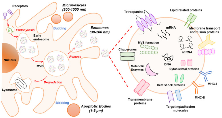
Communication between cells is crucial to preserve body homeostasis and health. Tightly controlled intercellular dialog is particularly relevant in the gut, where cells of the intestinal mucosa are constantly exposed to millions of microbes that have great impact on intestinal homeostasis by controlling barrier and immune functions. Recent knowledge involves extracellular vesicles (EVs) as mediators of such communication by transferring messenger bioactive molecules including proteins, lipids, and miRNAs between cells and tissues. The specific functions of EVs principally depend on the internal cargo, which upon delivery to target cells trigger signal events that modulate cellular functions. The vesicular cargo is greatly influenced by genetic, pathological, and environmental factors. This finding provides the basis for investigating potential clinical applications of EVs as therapeutic targets or diagnostic biomarkers. Here, we review current knowledge on the biogenesis and cargo composition of EVs in general terms. We then focus the attention to EVs released by cells of the intestinal mucosa and their impact on intestinal homeostasis in health and disease. We specifically highlight their role on epithelial barrier integrity, wound healing of epithelial cells, immunity, and microbiota shaping. Microbiota-derived EVs are not reviewed here.

Keywords: exosomes; extracellular vesicles; gut communication; gut immunity; intestinal homeostasis; miRNAs.


Conflict of interest statement

The authors declare no conflict of interest.

Figures

Figure 1
Figure 1
Schematic overview of extracellular vesicle (EV) biogenesis and cargo. At least three different subclasses of EVs are generated by eukaryotic cells: exosomes (30–200 nm), microvesicles (200–1000 nm), and apoptotic bodies (1–5 µm). The left panel schematically shows the biogenesis pathway for each EV type. Microvesicles and apoptotic bodies sprout directly from the plasma membrane, whereas exosomes are generated within multivesicular body (MVB) subpopulations that upon maturation fuse with the plasma membrane. The biogenesis pathway influences the cargo of EVs. In particular, the composition of exosomes is presented in the right panel. Exosomes are rich in the adhesion molecules tetraspanins (CD9, CD81, CD63), antigen-presenting molecules (MHCI/II), membrane transport proteins (annexins, flotillin), enzymes (elongation factors, metabolic enzymes), and other cytosolic proteins (ribosomal proteins). In addition, lipids (sphingomyelin and phosphatidylserine) and nucleic acids (DNA, RNA, non-coding RNAs (ncRNAs), and micro-RNAs (miRNAs)) also are bioactive components.
Figure 2
Figure 2
Modulation of immune responses by EVs originated from intestinal epithelial and immune cells. Schematic view of the intestinal mucosa showing the epithelium and the underlying immune system. These host cells receive information from microbiota mainly through secreted factors and lumen antigens that can diffuse through the mucus layer and initiate appropriate immune responses. In this scenario, EVs are key for the host to communicate with neighboring cells. Intestinal epithelial cells (IECs) release EVs from the basolateral side (colored in brown) that participate in the crosstalk with lymphocytes and dendritic cells, being able to activate naïve T cells towards immunogenic (Th) or tolerogenic (Treg) responses depending on the exosome-expressed epitopes and cargo. DCs are the main antigen-presenting cells in the gut lamina propria. These immune cells can integrate information either directly from the intestinal lumen or transmitted through EVs secreted by other cell types under healthy and pathological conditions (intestinal infections, inflammation, or cancer). Once activated, DCs secrete EVs (colored in purple) containing MHC and costimulatory molecules that mediate antigen presentation and immunomodulatory effects towards CD4+ or CD8+ T cells, eliciting suitable T cell responses.
Figure 3
Figure 3
Graphical summary of functions of EVs within the gut environment. EVs derived from IECs and immune cells of the lamina propria contribute to cell-to-cell communication in the gut and have great impact on the homeostasis/inflammation balance. The scheme summarized the bioactive cargo in EVs that can influence epithelial barrier integrity, tissue repair, immune responses, control of pathogens, and microbiota shaping.

Similar articles

Cited by

References

    1. Peterson L.W., Artis D. Intestinal epithelial cells: Regulators of barrier function and immune homeostasis. Nat. Rev. Immunol. 2014;14:141–153. doi: 10.1038/nri3608. - DOI - PubMed
    1. Yáñez-Mó M., Siljander P.R.M., Andreu Z., Zavec A.B., Borràs F.E., Buzas E.I., Buzas K., Casal E., Cappello F., Carvalho J., et al. Biological properties of extracellular vesicles and their physiological functions. J. Extracell. Vesicles. 2015;4:1–60. doi: 10.3402/jev.v4.27066. - DOI - PMC - PubMed
    1. Théry C., Witwer K.W., Aikawa E., Alcaraz M.J., Anderson J.D., Andriantsitohaina R., Antoniou A., Arab T., Archer F., Atkin-Smith G.K., et al. Minimal information for studies of extracellular vesicles 2018 (MISEV2018): A position statement of the International Society for Extracellular Vesicles and update of the MISEV2014 guidelines. J. Extracell. Vesicles. 2018;7:1–42. doi: 10.1080/20013078.2018.1535750. - DOI - PMC - PubMed
    1. Pegtel D.M., Gould S.J. Exosomes. Annu. Rev. Biochem. 2019;88:487–514. doi: 10.1146/annurev-biochem-013118-111902. - DOI - PubMed
    1. Zhang H., Freitas D., Kim H.S., Fabijanic K., Li Z., Chen H., Mark M.T., Molina H., Martin A.B., Bojmar L., et al. Identification of distinct nanoparticles and subsets of extracellular vesicles by asymmetric-flow field-flow fractionation. Nat. Cell Biol. 2018;20:332–343. doi: 10.1038/s41556-018-0040-4. - DOI - PMC - PubMed

MeSH terms

LinkOut - more resources