Review
. 2021 Feb 23;13(2):353.
doi: 10.3390/v13020353.

Recent Advances in Bunyavirus Glycoprotein Research: Precursor Processing, Receptor Binding and Structure

Affiliations
Review

Recent Advances in Bunyavirus Glycoprotein Research: Precursor Processing, Receptor Binding and Structure

Ruben J G Hulswit et al. Viruses. .

Abstract

The Bunyavirales order accommodates related viruses (bunyaviruses) with segmented, linear, single-stranded, negative- or ambi-sense RNA genomes. Their glycoproteins form capsomeric projections or spikes on the virion surface and play a crucial role in virus entry, assembly, morphogenesis. Bunyavirus glycoproteins are encoded by a single RNA segment as a polyprotein precursor that is co- and post-translationally cleaved by host cell enzymes to yield two mature glycoproteins, Gn and Gc (or GP1 and GP2 in arenaviruses). These glycoproteins undergo extensive N-linked glycosylation and despite their cleavage, remain associated to the virion to form an integral transmembrane glycoprotein complex. This review summarizes recent advances in our understanding of the molecular biology of bunyavirus glycoproteins, including their processing, structure, and known interactions with host factors that facilitate cell entry.

Keywords: Bunyavirales; bunyavirus; genomic coding strategy; glycoprotein precursor processing; glycoproteins; protein structure; receptor binding.


Conflict of interest statement

The authors declare no conflict of interest.

Figures

Figure 1
Figure 1
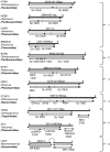
Families belonging to the Bunyavirales order. Polymerase sequences of a representative set of bunyaviruses were aligned using MAFFT (Multiple Alignment using Fast Fourier Transform) [20], the alignment was trimmed using trimAI [21] and a phylogenetic tree created in PhyML [22] using the Smart Model Selection option [23]. The unrooted tree was displayed using iTOL (interactive Tree of Life) [24]. Asterisks indicate main branches for which bootstrap support was <0.7. The dataset used for the phylogeny includes all currently assigned bunyavirus families [1], the grouping of which is broadly in line with that presented in a recently published tree [25]. The numbered, black lines in the figure denote bunyavirus species currently not assigned to a family [Wuhan spider virus (1), Laurel lake virus (2), Hubei blood fluke virus 2 (3), Hubei blood fluke virus 1 (4), Mothra virus (5), Shistocephalus solidus bunya-like virus (6)]. Common organisms from which the viruses were isolated are indicated by symbols and viruses known to be capable of zoonotic transmission to humans are annotated with red symbols (the panel at the right denotes what the symbols represent).
Figure 2
Figure 2
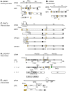
Genomic structure and coding strategies for the viral genomic RNA segments coding for viral glycoprotein precursors (GPCs) from well-studied families within the Bunyavirales order. Genomic RNAs (3’ to 5’) are represented by black lines (the numbers of nucleotides and the amino acid of GPC are given above). mRNAs are shown as arrows (♦ indicates host-derived primer sequence at 5’ end by cap-snatching mechanism). Gene products are presented by bars with their approximate size shown underneath. The sites for cleavage are represented by “|”. Virus abbreviations: HTNV, Hantaan virus; HEBV, Herbet virus; UUKV, Uukuniemi virus; EMARaV, European mountain ash ringspot-associated virus; BUNV, Bunyamwera virus; RVFV, Rift Valley fever virus; FERV, Fermo virus; CCHFV, Crimean-Congo haemorrhagic fever virus; TSWV, Tomato spotted wilt virus; RSV, rice stripe virus; LASV, Lassa virus. (1) The M segments of hantaviruses (Hantaviridae) [33], herbeviruses (Peribunyaviridae) [34], Uukuviruses (e.g., UUKV) (Phenuiviridae) [35], and the RNA2 segment of the emaraviruses (Fimoviridae) [36] encode GPCs containing two structural glycoproteins, Gn and Gc. (2) The M segments of orthobunyaviruses (Peribunyaviridae) [11] encode three proteins, with an NSm located between Gn and Gc in the precursor protein. (3) The M segments of phleboviruses (e.g., RVFV) (Phenuiviridae) [37] and orthophasmaviruses (e.g., Ferak virus [FRKV] and jonchet virus, [JONV] (Phasmaviridae) encode GPCs containing three proteins: Gn and Gc, and an N-terminal NSm [38,39]. (4) The M segment of nairoviruses (e.g., CCHFV) (Nairoviridae) encodes a GPC with five proteins: Gn and Gc, and three non-structural proteins; Mucin like protein/domain (MLD), GP38, and NSm [40,41]. The M segments of other members of the family encode precursors for two to four proteins whose exact nature has yet to be confirmed experimentally [42]. (5) The ambisense M segments of orthotospoviruses (Tospoviridae) [29] and RNA2 segments of tenuiviruses (Phenuiviridae) encode GPCs containing Gn and Gc in the antigenomic sense, and an NSm in the genomic sense [43]. (6) The ambisense S segments of members of the Hartmanivirus, Mammarenavirus and Reptarenavirus genera (Arenaviridae family) encode a so-called stable signal peptide (SSP) and the glycoproteins GP1 and GP2 in the genomic sense, and a nucleocapsid protein in the antigenomic sense [26]. Similarly, the M segment of Wēnlǐng frogfish arenaviruses (Antennavirus genus) encode their GPCs in the genomic sense and an unknown protein in the anti-genomic sense [44].
Figure 3
Figure 3
Current models of bunyaviral GPC processing for selected peribunyaviruses, hantaviruses, phenuiviruses, nairoviruses, and arenaviruses. GPC polyproteins are shown for BUNV (A), HTNV (B), RVFV (C), CCHFV (D) and LASV (E). The signal peptide (SP) and stable SP (SPP, for LASV) are represented in orange. Transmembrane domains (TM) are shown as black boxes. The cleavage sites for signal peptidase (SPase) and signal peptide peptidase (SPP) are marked by scissors and an arrowhead (➣), respectively. The SKI-1/S1P cleavage site is marked by a downward arrow. Nonstructural proteins (NSm and GP38) are shown as grey boxes and the CCHFV mucin domain as a grey and white box. The pre-Gc N-terminal 41 residues that are processed at the RKPR/RKPL motif by a SKI-1/S1P-like protease are shown as green box.
Figure 4
Figure 4
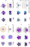
Envelope glycoprotein ultrastructure of orthobunya-, hanta-, phlebo- and arenavirus particles. (A) Left panel: an EM reconstruction of an orthobunyaviral envelope glycoprotein (in grey; EMD-23527) obtained using Bunyamwera (BUNV) particles, to which the trimeric BUNV Gc head domain structure (PDB: 6H3V8) was fitted, in side view. The center of the tripodal organization is linked together by the membrane-distal, N-terminal extension of the Gc. The different regions of the Gn-Gc spike complex (head, stalk, floor) are indicated on the left with corresponding heights. The head and stalk regions make up the N-terminal extension of the peribunyaviral Gc glycoprotein, whereas the floor region is thought to represent the Gn ectodomain and the fusogenic C-terminal region of the Gc ectodomain. Middle panel: zoomed-in surface representation of the Gc head domain trimer. Each of the protomers is colored in a different shade of blue. Right panel: Ribbon presentation of the middle panel. (B) Top views of the three upper panels. Left panel: the different threefold symmetry axes are indicated by open or solid triangles. (C) Left panel: EM density corresponding to the region of a phleboviral envelope Gn-Gc complex that exhibits five-fold symmetry (in grey; EMD-4201 [151]), obtained using Rift Valley fever virus (RVFV) particles. RVFV Gn and Gc crystal structures were fitted into this this pentameric RVFV envelope glycoprotein capsomer (PDB: 6F9F [151]), in side view. N-terminal regions of the Gn (membrane distal) are indicated in shades of purple and pink and the Gc ectodomain (membrane proximal) in shades of blue. Glycoprotein EM density is shown in grey. Middle panel: surface representation of the pentameric organization of Gn/Gc heterodimers. Right panel: Ribbon presentation of the middle panel. (D) Top views of the three upper panels. Left panel: the fivefold symmetry axis is indicated by a solid pentagon. (E) Left panel: the EM density of a hantaviral envelope Gn-Gc complex (in grey; EMD-11236, [154]) obtained using Tula virus (TULV) particles to which a model of tetrameric (Gn-Gc)4 ANDV spike (PDB: 6ZJM, [154]) was fitted, in side view. ANDV Gn is indicated in shades of purple and pink and ANDV Gc in shades of blue. Middle panel: surface representation of the tetrameric organization of Gn/Gc heterodimers. Right panel: ribbon presentation of the middle panel. (F) Top views of the three upper panels. Left panel: the fourfold symmetry axis is indicated by a solid square. (G) Left panel: EM density of a trimeric arenavirus envelope glycoprotein spike (in grey; EMD-3290 [155]) obtained using Lassa virus (LASV) particles to which the trimeric LASV GP1-GP2 envelope glycoprotein (PDB: 5VK2 [156]) was fitted, in side view. GP1 protomers (membrane distal) are indicated in shades of purple and pink and GP2 protomers (membrane proximal) in shades of blue. Glycoprotein EM density is shown in grey. Middle panel: zoomed-in surface representation of the trimeric organization of LASV GP1/GP2 heterodimers. Right panel: Ribbon presentation of the middle panel. (H) Top views of the three upper panels. Left panel: the threefold symmetry axis is indicated by a solid triangle. In all structural representations crystallographically observed glycans are shown as white sticks. In the case of the RVFV Gn-Gc pentameric assembly, glycan chains were modelled onto N-linked glycosylation sites. The position of the viral membrane is shown as a yellow surface or two yellow dashed lines. To emphasize the protein components of the reconstructions, lipid bilayer EM densities were rendered at a lower sigma level than the protein surfaces. A color legend is shown on the top right-hand side of each panel.
Figure 5
Figure 5
Known structural features of bunyaviral Gn and Gc envelope proteins. (A) The Gn envelope glycoprotein displays limited structural similarity across bunyavirus families. Five crystal structures of Gn ectodomain regions of different bunyaviruses are shown. Upper panel: cartoon representation of the Gn N-terminal region of the ectodomain of the New World orthohantavirus ANDV (PDB: 6Y6P [154]), which displays a four-domain architecture (domain A, deep purple; a β-ribbon domain, purple-brown; domain B, warm pink; and a domain reminiscent of the alphavirus E3 protein, white). Second panel: cartoon representation of the Gn N-terminal region of the ectodomain of the Old World orthohantavirus PUUV (PDB: 5FXU [165]). Third panel: cartoon representation of the N-terminal region of the Gn ectodomain from RVFV (PDB: 6F8P [151]). Fourth panel: cartoon representation of the N-terminal region of the Gn ectodomain from SFTSV (PDB: 5Y10 [166]). Interestingly, SFTSV Gn contains a region reminiscent of the E3-like domain observed in hantavirus Gn proteins. Bottom panel: cartoon representation of the Gn ectodomain from TSWV (PDB: 6Y9L [167]). TSWV Gn displays a largely conserved three-domain architecture in which domain B is reduced to a β-hairpin. The C-terminal domain (CTD) comprises a β-sandwich domain (cyan) (please see Figure 6). (B) Structurally characterized bunyaviral Gc fusion proteins display a conserved class II fusion protein architecture (domain I, light blue; domain II, slate blue; domain III, sky blue). Four crystal structures of the Gc ectodomain of different bunyaviruses are shown in a putative pre-fusion conformation (except SFTSV Gc for which a post-fusion state was determined). The dashed grey arrow indicates the movement of domain III between putative pre- and post-fusion conformations. Fusion loop(s) are indicated in bright orange. Top panel: crystal structure of the ANDV New World orthohantavirus Gc protein ectodomain in its pre-fusion conformation (PDB: 6Y5F [154]). Second panel: crystal structure of the Old World orthohantavirus PUUV Gc protein ectodomain in its pre-fusion conformation (PDB: 7B09 [168]). Third panel: crystal structure of the RVFV phlebovirus Gc protein ectodomain in its pre-fusion conformation (PDB: 4HJ1 [160]). Bottom panel: crystal structure of the SFTSV Gc protein ectodomain in its post-fusion conformation (PDB: 5G47 [169]). Note that the position of domain III has shifted from the tip of domain I in pre-fusion conformations towards the border of domains I and II in this post-fusion state. In all structural representations crystallographically observed glycans are shown as white sticks.
Figure 6
Figure 6
The C-terminal regions of ANDV Gn, TSWV Gn, and alphavirus E2 ectodomains have been shown to contain a seven-stranded β-sandwich fold. Rainbow cartoon representation of the aligned C-terminal β-sandwich domains of ANDV Gn, TSWV Gn and CHIKV E2. (A) The N-terminus of the displayed ANDV Gn structure (PDB: 6YRB, [154]) connects to the multi-domain globular region presented in Figure 5 and the C-terminus connects to an α-helical hairpin (grey cartoon), also known as the ‘base’ domain. (B) The N-terminus of the displayed TSWV Gn structure (PDB: 6Y9L, [167]) connects to the multi-domain globular region (grey cartoon, also presented in Figure 5) and the C-terminus connects to the transmembrane domain. (C) The N-terminus of the displayed CHIKV E2 structure (PDB: 3N43, [170]) connects to a multi-domain globular region (partially shown by grey cartoon) and the C-terminus connects to the transmembrane domain. The N- and C-termini of the β-sandwich folds are colored blue and red, respectively, and the N- and C-termini of the structures are indicated.
Figure 7
Figure 7
Crystal structures of the orthobunyavirus Gc N-terminal region, CCHFV GP38, and arenavirus glycoproteins. (A) Crystal structure of the multi-domain N-terminal region of SBV Gc (head domain, light blue; stalk domain I, violet purple; stalk domain II, blue white) (PDB: 6H3S [153]). The N- terminus of the protein is indicated, as is the C-terminus that connects to the Gc protein. (B) Crystal structure of CCHFV GP38 protein (PDB: 6VKF [175]). The N- and C-termini of the protein are indicated. (C) Single protomer of the trimeric LASV GP crystal structure (PDB: 5VK2 [156]). The N- and C-termini of the GP1 and GP2 components are indicated. (D) Crystal structure of the trimeric post-fusion state of LASV GP2 (PDB: 5OMI [176]). N- and C-termini of the different GP2 protomers are indicated. In all structural representations crystallograhpically observed glycans are shown as white sticks.
Figure 8
Figure 8
Diverse class II fusion protein architectures converge on an evolutionary conserved trimeric post-fusion conformation. Schematic representation of class II fusion proteins in their pre-fusion oligomeric state. The schematic assemblies are based on crystal structures and/or cryoEM reconstructions (Peribunyaviridae: BUNV (EMD-2352 [163]) and SBV (PDB: 6H3V [153]); Hantaviridae: TULV (EMD-3364 [165]); Phenuiviridae: RVFV (EMD-4201 and PDB: 6F9F [151]); Flaviviridae: (PDB: 4UTC [189]); Togaviridae: Alphavirus eastern equine encephalitis virus (EEEV; PDB: 6MX4 [190]), Rubivirus RUBV (PDB: 4ADJ[191]); C. elegans (PDB: 4OJC [149]). The elongated structures of class II fusion proteins are shown as blue shapes (Gc for members of the Bunyavirales, E1 for Togaviridae, E for Flaviviridae and EFF-1 for the cellular C. elegans protein). Putative fusion protein stabilizing entities present on mature viral particles, are shown as purple shapes and have been hypothesized to prevent premature fusion activation (Bunyavirales: Gn, Togaviridae: E2). The E3 protein has been shown to be present in some alphavirus particles [170] but is omitted from this representation for clarity. The level of symmetry of each of the protein assemblies is indicated by symmetry symbols at the bottom right-hand corner. The approximate position of the fusion loop(s) is indicated with an asterisk (*) for each panel. In the case of peribunyaviruses, the exact location of the fusion loop (white asterisk) within the Gc protein is currently not known, but was inferred from the location of the N-terminal extensions within the tripodal EM reconstruction [153,163] and the C-terminal positioning of Gc transmembrane domains. Note that, although C. elegans EFF-1 (epithelial fusion failure 1) protein presents a class II fusogen architecture, it does not contain a fusion loop. Fusion is believed to be initiated by trimerization of the plasma membrane anchored EFF-1 ectodomains protruding in the extracellular space [149]. The grey region of the column shown for Peribunyaviridae represents the N-terminal extension of the Gc fusion protein, which has not been observed in other bunyavirus glycoproteins. The pre-fusion oligomeric state of EFF-1 has been observed to be monomeric on the plasma membrane [192]. The pre-fusion oligomeric state of rubella virus E1 on the virus membrane is currently unknown and therefore represented as a protomer of an unknown oligomeric assembly. The fusion proteins of alpha- (e.g., Semliki Forest virus (SFV), chikungunya virus (CHIKV)) and flaviviruses (e.g., dengue virus (DENV), zika virus (ZIKV)) are structurally related despite a lack of detectable sequence conservation and are therefore positioned next to each other in the diagram. Similarly, phenuivirus Gc has been shown to be genetically more closely related to the fusion envelope (E) proteins of flaviviruses than to those of other genera in its own order [152]. These proteins are placed next to each other to represent this predicted relationship. The box depicting the cellular EFF-1 protein is colored in yellow as to oppose the boxes in different shades of blue which all contain viral fusion proteins.

Similar articles

Cited by

References

    1. Abudurexiti A., Adkins S., Alioto D., Alkhovsky S.V., Avšič-Županc T., Ballinger M.J., Bente D.A., Beer M., Bergeron É., Blair C.D., et al. Taxonomy of the order Bunyavirales: Update 2019. Arch. Virol. 2019;164:1949–1965. doi: 10.1007/s00705-019-04253-6. - DOI - PMC - PubMed
    1. Kuhn J.H., Adkins S., Alioto D., Alkhovsky S.V., Amarasinghe G.K., Anthony S.J., Avšič-Županc T., Ayllón M.A., Bahl J., Balkema-Buschmann A., et al. 2020 taxonomic update for phylum Negarnaviricota (Riboviria: Orthornavirae), including the large orders Bunyavirales and Mononegavirales. Arch. Virol. 2020;165:3023–3072. doi: 10.1007/s00705-020-04731-2. - DOI - PMC - PubMed
    1. Mehand M.S., Al-Shorbaji F., Millett P., Murgue B. The WHO R&D blueprint: 2018 review of emerging infectious diseases requiring urgent research and development efforts. Antivir. Res. 2018;159:63–67. - PMC - PubMed
    1. Plyusnin A., Beaty B.J., Elliott R.M., Kormelink R., Lundkvist A., Schmaljohn C.S., Tesh R.B. Bunyaviridae. In: Andrew M.Q., King M.J.A., Eric B.C., Lefkowitz E.J., editors. Virus Taxonomy. Elsevier; Oxford, UK: 2011. pp. 725–742.
    1. Elliott R.M., Schmaljohn C.S. Bunyaviridae. In: Knipe D.M., Howley P.M., editors. Fields Virology. 6th ed. Wolters Kluwer; Philadelphia, PA, USA: 2013. pp. 1244–1282.

Publication types

MeSH terms