. 2021 Feb 23;13(2):355.
doi: 10.3390/v13020355.

Integrase-Defective Lentiviral Vector Is an Efficient Vaccine Platform for Cancer Immunotherapy

Affiliations

Integrase-Defective Lentiviral Vector Is an Efficient Vaccine Platform for Cancer Immunotherapy

Valeria Morante et al. Viruses. .

Abstract

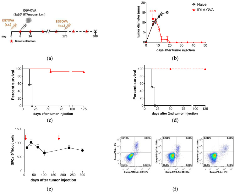
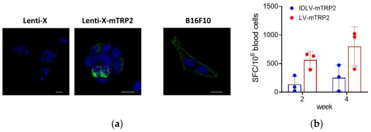
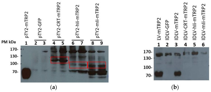
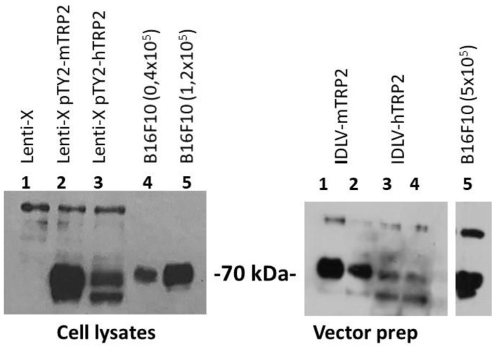
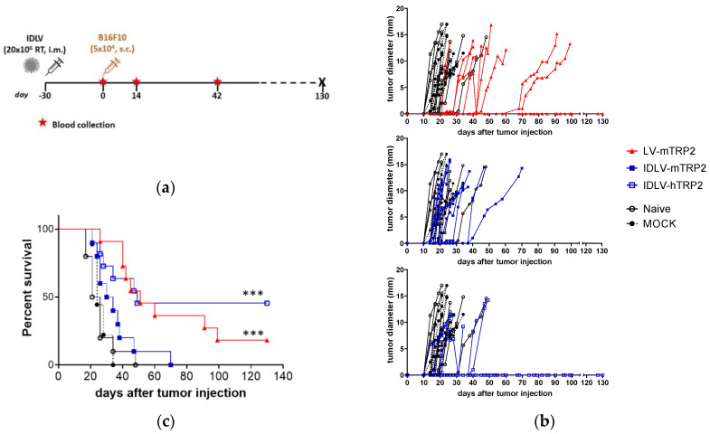
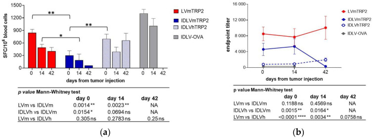
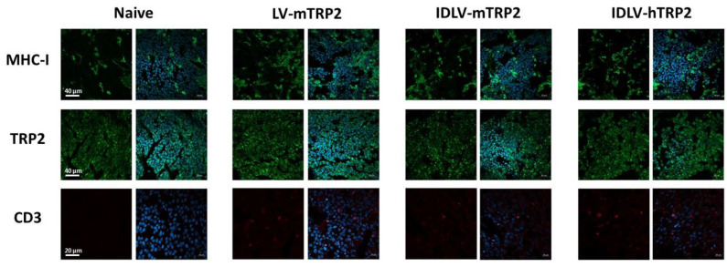
Integrase-defective lentiviral vectors (IDLVs) have been used as a safe and efficient delivery system in several immunization protocols in murine and non-human primate preclinical models as well as in recent clinical trials. In this work, we validated in preclinical murine models our vaccine platform based on IDLVs as delivery system for cancer immunotherapy. To evaluate the anti-tumor activity of our vaccine strategy we generated IDLV delivering ovalbumin (OVA) as a non-self-model antigen and TRP2 as a self-tumor associated antigen (TAA) of melanoma. Results demonstrated the ability of IDLVs to eradicate and/or controlling tumor growth after a single immunization in preventive and therapeutic approaches, using lymphoma and melanoma expressing OVA. Importantly, LV-TRP2 but not IDLV-TRP2 was able to break tolerance efficiently and prevent tumor growth of B16F10 melanoma cells. In order to improve the IDLV efficacy, the human homologue of murine TRP2 was used, showing the ability to break tolerance and control the tumor growth. These results validate the use of IDLV for cancer therapy.

Keywords: immune tolerance; immunotherapy; lentiviral vector: vaccine; tumor; tumor associated antigen.


Conflict of interest statement

The authors declare no conflict of interest.

Figures

Figure 1
Figure 1
Therapeutic efficacy of integrase-defective lentiviral vectors (IDLV)-ovalbumin (OVA) vaccination in E.G7-OVA-bearing mice. (a) Scheme of the experiment: 14 C57BL/6 mice were inoculated s.c. with 3.5 × 10⁶ E.G7-OVA cells/mouse. Mice with 7–9 mm diameter tumor mass were vaccinated with 3 × 10⁶ RT/mouse of IDLV-OVA (n = 7) or left untreated (Naïve, n = 7). Tumor growth, survival, and immune response were monitored over time. Tumor-free mice were injected a second time with tumor cells at 175 days and monitored up to the end of the experiment (300 days). (b) Tumor growth after the first tumor injection is shown. Mice were sacrificed when the tumor diameter reached 15 mm or an ulceration of tumor was observed. (c,d) Kaplan–Meier survival curves are shown. Survival was monitored up to 175 days after the first tumor injection (Log-rank Mantel-Cox test) and up to 125 days from the second tumor injection (Log-rank Mantel-Cox test). (e) Kinetics of OVA-specific T cell response in IDLV-OVA vaccinated mice, after E.G7-OVA injection (red arrows). Blood cells were collected at the indicated time points and stimulated with the H-2Kb restricted OVA 8mer peptide (SIINFEKL). Data are expressed as specific spot forming cells (SFC) per 106 cells. Error bars indicate the standard deviation among the animals from the same group. (f) Polyfunctional OVA-specific CD8+ T cells. Vaccinated and tumor-free mice were sacrificed at 300 days and splenocytes were used to evaluate the magnitude and quality of OVA-specific CD8+ T cell response by intracellular cytokine staining (ICS). A representative experiment is shown. CD8+ T cells were analyzed in splenocytes stimulated with OVA 8mer peptide (OVApep). The percentage of CD8+ T cells producing IFNγ and TNFα and expressing CD107a is indicated within the quadrants.
Figure 2
Figure 2
Therapeutic efficacy of IDLV-OVA vaccination in B16OVA-bearing mice. (a) Scheme of the experiment. C57BL/6 mice were s.c. injected with 2 × 10⁵ B16OVA cells/mouse. After 12 days, groups of mice were vaccinated either with 10 × 106 RT/mouse of IDLV-OVA or IDLV expressing an unrelated antigen (mock) or left untreated (Naïve). (b) OVA-specific T cell response was generated in all IDLV-OVA immunized mice, as evaluated by IFNγ ELISpot measured two weeks after immunization in blood cells (left panel) and at sacrifice in splenocytes (right panel). Cells were collected and stimulated with H-2Kb restricted OVA 8mer peptide (SIINFEKL). Data are expressed as spot forming cells (SFC) per million cells. (c) Tumor growth. All groups developed a tumor mass measured until the end of the experiment. The delay of tumor growth in IDLV-OVA immunized mice is highlighted with the red rectangle. Mock and Naïve groups were sacrificed within 18 days from tumor injection. (d) Kaplan–Meier survival curve. Mice with tumor diameter >15 mm or a serious ulceration were sacrificed. (Log-rank Mantel-Cox test, **** p < 0.0001).
Figure 3
Figure 3
Confocal laser scanning microscopy (CLSM) analyses on mice tissue sections. Representative images of tumor from Naïve and IDLV-OVA mice are shown. Tissue sections 8 µm thick were stained for MHC Class I (green), OVA (green), or CD3 (red) as indicated (left columns) and for DAPI as nuclear staining (blue, right columns). Images represent a 3D reconstruction of 30–40 single Z-stack. Results from one representative experiment are shown for each analysis. Scale bars are 40 µm.
Figure 4
Figure 4
IDLV expressing mTRP2 as a self-antigen induces low specific T cells. (a) Expression of mTRP2 in cell lines. Mock Lenti-X cells, pTY2CMVmTRP2W-transfected Lenti-X cells and B16F10 cells were fixed and stained with anti-TRP2 antibody (green) and DAPI (blue) and analyzed by CLSM. Images represent a 3D reconstruction from 14 single optical sections. Results from one representative experiment are shown for each analysis. Scale bars are shown for each figure. (b) Pilot study of immunogenicity. C57Bl6 mice (n = 3) were immunized once with 10 × 106 RT units/mouse of either IDLV-mTRP2 or LV-mTRP2. The TRP2-specific T cell-mediated immune response was evaluated by IFNγ-ELISpot assay at two and four weeks in blood. Blood cells were stimulated with H-2Kb restricted TRP2 9mer peptide (SVYDFFVWL). Data are expressed as specific spot forming cells (SFC) per million cells. Box plots show mean ± SEM and single values from each immunized mouse.
Figure 5
Figure 5
Expression of different TRP2 fusion proteins by Western blot. (a) Cell lysates of Lenti-X transfected with transfer vector expressing either mTRP2 (predicted size 70 kDa) or mTRP2 fused with CRT (Calreticulin, predicted size 123 kDa), hIi (human invariant chain, predicted size 109 kDa), or mIi (murine invariant chain, predicted size 106 kDa). Lenti-X cells transfected with mTRP2 wild type (5 × 10⁴ cells, lane 1) and GFP transfer vectors (lanes 2 and 3) were used as positive and negative control, respectively. The assay was performed using 1.6 × 10⁵ (lane 2–4–6–8) or 3.2 × 10⁵ cells (lane 3–5–7–9). Red boxes indicate the band with the correct molecular weight. (b) Detection of mTRP2 in viral vector preparations. 1.5 × 10⁶ RT units of LV-mTRP2 (lane 1), IDLV-GFP (lane 2), IDLV-mTRP2 (lane 3), IDLV-CRT-mTRP2 (lane 4), IDLV-hIi-mTRP2 (lane 5), IDLV-mIi-mTRP2 (lane 6) were analyzed.
Figure 6
Figure 6
Expression of murine and human TRP2 evaluated by Western blot. Left panel: Detection of TRP2 in cell lysates of Lenti-X cells (1 × 10⁵) transfected with plasmids expressing either mTRP2 or hTRP2. Lenti-X and B16F10 cell lysates were used as negative and positive control of TRP2 expression, respectively. Right panel: Detection of TRP2 in concentrated vector preparations (3 × 10⁶ RT/lane). The rabbit anti-TRP2 polyclonal antibody used to detect the expression of TRP2 recognizes both human and murine TRP2 proteins.
Figure 7
Figure 7
Antitumor efficacy of lentiviral vectors expressing either murine or human TRP2. (a) Scheme of the experiment. C57BL/6 mice (n = 9–11) were immunized with vectors expressing TRP2, OVA (Mock) or left untreated. After 30 days all mice were s.c. injected with 5 × 10⁴ B16F10 cells/mouse. (b) Tumor growth was monitored over time. All groups developed a tumor mass measured until the end of the experiment. Mock and Naïve groups were sacrificed within 32 days from tumor injection. (c) Survival was monitored over time and Kaplan–Meier survival curve is shown. Mice with tumor diameter >15 mm or a serious ulceration were sacrificed. (Log-rank Mantel-Cox test, *** p < 0.0001).
Figure 8
Figure 8
Kinetics of TRP2-specific immune responses in mice immunized with IDLV delivering either murine or human TRP2 and challenged with B16F10 tumor. C57BL/6 mice (n = 9–11) immunized with vectors expressing TRP2, OVA, or left untreated were s.c. injected with 5 × 10⁴ B16F10 cells/mouse after 30 days from immunization, as depicted in Figure 7a. (a) TRP2-specific T cell response was evaluated by IFNγ ELISPOT, measured at different time points in blood. Cells were collected and stimulated with H-2Kb restricted TRP2 9mer peptide (SVYDFFVWL). Data are expressed as mean spot forming cells (SFC) per 106 cells, bars represent standard error among animals from the same group. (b) Anti-mTRP2 IgG antibodies were analyzed in plasma of immunized animals by ELISA. Data are expressed as mean of endpoint titers and bars represent standard error among animals from the same group. Comparison among groups was evaluated using the Mann–Whitney test, as indicated by p values shown in the tables under the graphs. * p < 0.05; ** p < 0.01; **** p < 0.0001. NA: The comparison was not performed since only one animal from IDLVmTRP2 group was alive at day 42.
Figure 9
Figure 9
Confocal laser scanning microscopy (CLSM) analyses on mice tissue sections. Representative images of tumor from naïve, LV-mTRP2, IDLV-mTRP2, and IDLV-hTRP2 mice are shown. Tissue sections 8 µm thick were stained for MHC–I (green), TRP2 (green), or CD3 (red) as indicated (left columns) and for DAPI as nuclear staining (blue, right columns). Images represent a 3D reconstruction of 30–40 single Z-stack. Results from one representative experiment are shown for each analysis. Scale bars are indicated.

References

    1. FUTURE II Study Group Quadrivalent vaccine against human papillomavirus to prevent high-grade cervical lesions. N. Engl. J. Med. 2007;356:1915–1927. doi: 10.1056/NEJMoa061741. - DOI - PubMed
    1. Kantoff P.W., Higano C.S., Shore N.D., Berger E.R., Small E.J., Penson D.F., Redfern C.H., Ferrari A.C., Dreicer R., Sims R.B., et al. IMPACT Study Investigators. Sipuleucel-T immunotherapy for castration-resistant prostate cancer. N. Engl. J. Med. 2010;363:411–422. doi: 10.1056/NEJMoa1001294. - DOI - PubMed
    1. Maude S.L., Frey N., Shaw P.A., Aplenc R., Barrett D.M., Bunin N.J., Chew A., Gonzalez V.E., Zheng Z., Lacey S.F., et al. Chimeric antigen receptor T cells for sustained remissions in leukemia. N. Engl. J. Med. 2014;371:1507–1517. doi: 10.1056/NEJMoa1407222. - DOI - PMC - PubMed
    1. Wagner J., Wickman E., DeRenzo C., Gottschalk S. CAR T Cell Therapy for Solid Tumors: Bright Future or Dark Reality? Mol. Ther. 2020;28:2320–2339. doi: 10.1016/j.ymthe.2020.09.015. - DOI - PMC - PubMed
    1. Hodi F.S., O’Day S.J., McDermott D.F., Weber R.W., Sosman J.A., Haanen J.B., Gonzalez R., Robert C., Schadendorf D., Hassel J.C., et al. Improved survival with ipilimumab in patients with metastatic melanoma. N. Engl. J. Med. 2010;363:711–723. doi: 10.1056/NEJMoa1003466. - DOI - PMC - PubMed

Publication types

MeSH terms

LinkOut - more resources