. 2021 Feb 23;11(2):557.
doi: 10.3390/nano11020557.

The Effect of Selenium Nanoparticles on the Osteogenic Differentiation of MC3T3-E1 Cells

Affiliations

The Effect of Selenium Nanoparticles on the Osteogenic Differentiation of MC3T3-E1 Cells

Sang-Cheol Lee et al. Nanomaterials (Basel). .

Abstract

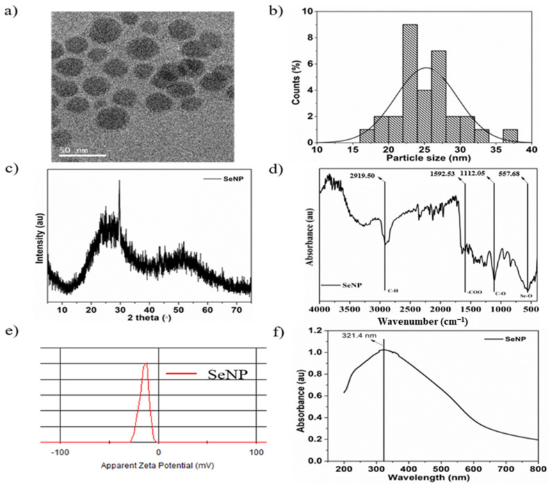
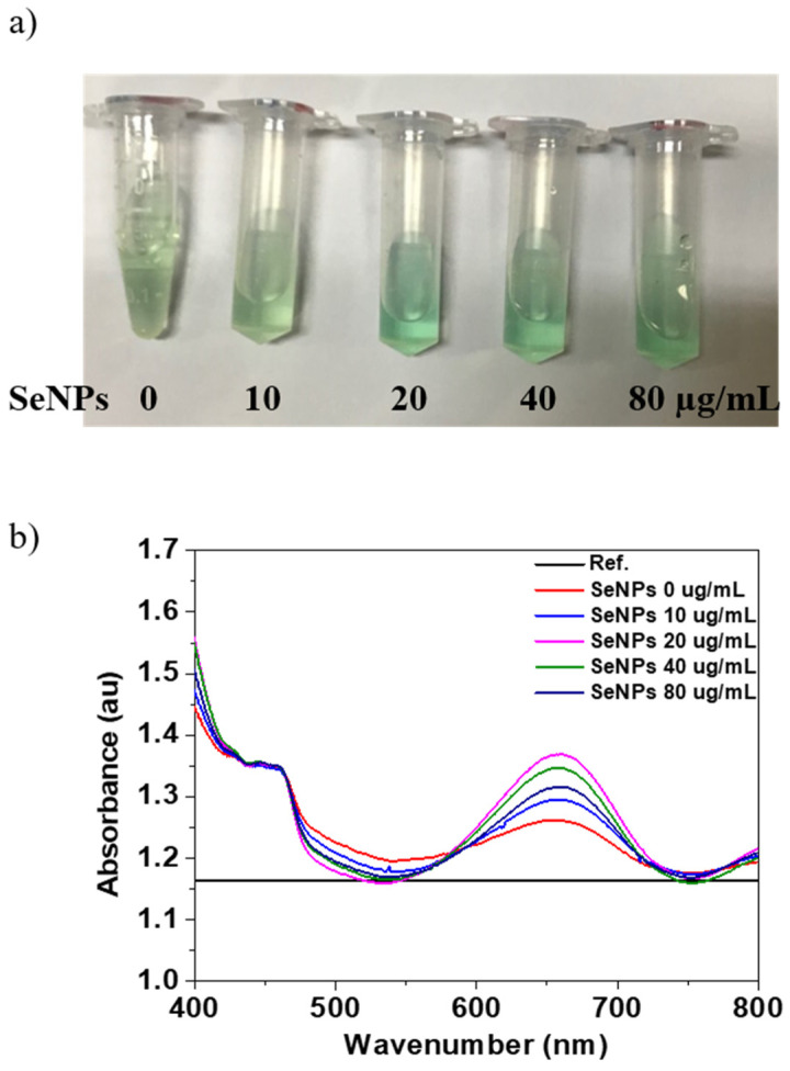
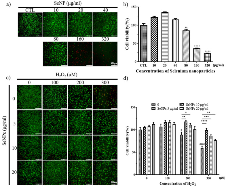
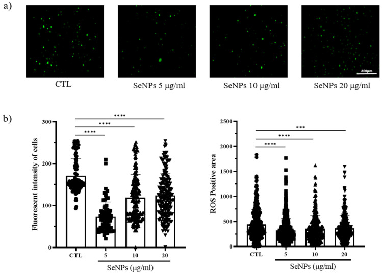
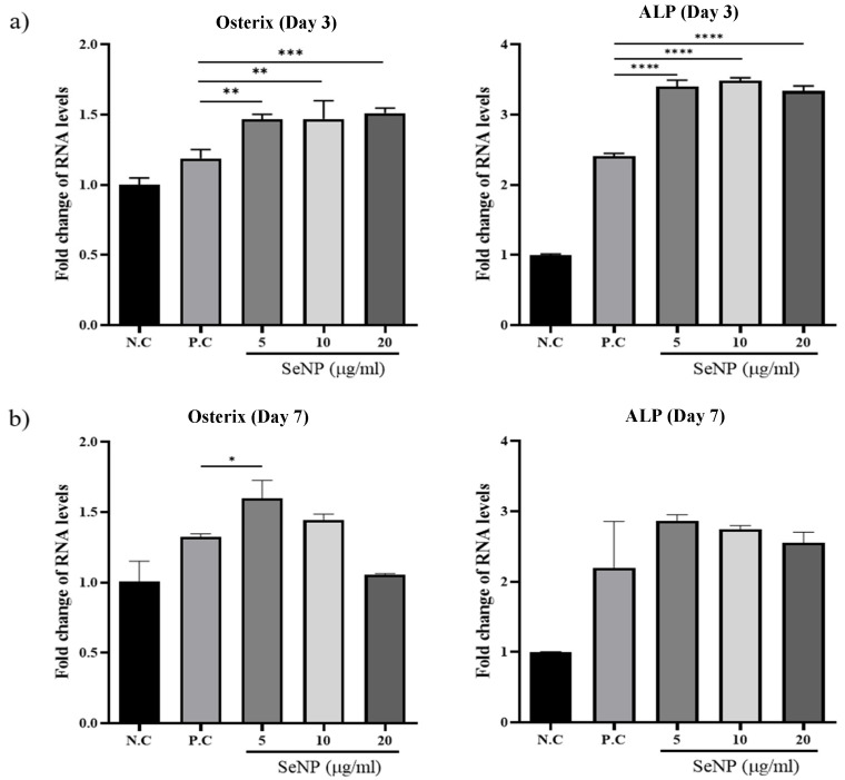
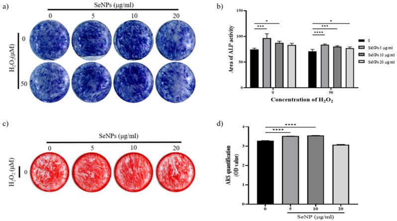
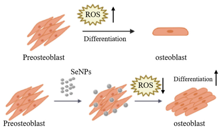
Reactive oxygen species (ROS) regulate various functions of cells, including cell death, viability, and differentiation, and nanoparticles influence ROS depending on their size and shape. Selenium is known to regulate various physiological functions, such as cell differentiations and anti-inflammatory functions, and plays an important role in the regulation of ROS as an antioxidant. This study aims to investigate the effect of selenium nanoparticles (SeNPs) on the differentiation of osteogenic MC3T3-E1 cells. After fabrication of SeNPs with a size of 25.3 ± 2.6 nm, and confirmation of its oxidase-like activity, SeNPs were added to MC3T3-E1 cells with or without H2O2: 5~20 μg/mL SeNPs recovered cells damaged by 200 μM H2O2 via the intracellular ROS downregulating role of SeNPs, revealed by the ROS staining assay. The increase in osteogenic maturation with SeNPs was gradually investigated by expression of osteogenic genes at 3 and 7 days, Alkaline phosphatase activity staining at 14 days, and Alizarin red S staining at 28 days. Therefore, the role of SeNPs in regulating ROS and their therapeutic effects on the differentiation of MC3T3-E1 cells were determined, leading to possible applications for bone treatment.

Keywords: MC3T3-E1; nanoparticles; osteogenic differentiation; reactive oxygen species; selenium.


Conflict of interest statement

The authors declare no conflict of interest. The funders had no role in the design of the study; in the collection, analyses, or interpretation of data; in the writing of the manuscript, or in the decision to publish the results.

Figures

Figure 1
Figure 1
Characterization of selenium nanoparticles (SeNPs): (a) Transmission electron microscopy (TEM) image of SeNPs, (b) particle size distribution, (c) X-ray diffraction (XRD) pattern, (d) Fourier transform infrared (FTIR) spectrum, (e) ξ-potential, and (f) UV-visible spectrum of SeNPs in distilled water.
Figure 2
Figure 2
Determination of oxidase properties of different concentrations of SeNPs: (a) The color evaluation of TMB oxidation with SeNP concentrations of 0, 10, 20, 40, and 80 µg/mL, (b) the UV-visible absorption spectra of TMB (before reaction as Reference) and after oxidation for 30 min with and without SeNPs.
Figure 3
Figure 3
Cell viability and proliferation were determined by live (green) and dead (red) and CCK-8 assays. After cell seeding for 24 h, MC3T3-E1 cells were analyzed using CCK-8 solution and stained using live and dead staining to evaluate cell viability. (a) Live and dead staining following treatment with various SeNP concentrations. (b) The relative cell viability of MC3T3-E1 cells cultured in different concentrations of SeNPs. (c) Live and dead staining following treatment with various SeNP concentrations and H2O2. (d) Cell viability of MC3T3-E1 cells treated with various concentrations of SeNPs and H2O2. The statistical significance of (b) was calculated using one-way analysis of variance (ANOVA), and (d) was calculated using two-way ANOVA followed by a two-sided Dunnett’s multiple comparison test compared to control (CTL) (scale bar = 350 μm). * Represents p < 0.05, ** p < 0.01, *** p < 0.001, **** p < 0.0001, # is compared with SeNPs and H2O2-untreated groups. # Represents p < 0.05, #### p < 0.0001; n = 4.
Figure 4
Figure 4
MC3T3-E1 cells were exposed to 400 μM H2O2 for oxidative stress and then recovered by culturing in medium with or without SeNPs. High oxidative stress conditions were enabled by pretreatment with H2O2 for 4 h. (a) SeNP treatment resulted in reduced levels of reactive oxygen species (ROS). (b) The fluorescence intensity of cells and ROS-positive areas was measured using ImageJ. Statistical significance was calculated using one-way ANOVA followed by a two-sided Dunnett post hoc test compared to CTL (scale bar = 350 μm). **** Represents p < 0.0001; n = 5.
Figure 5
Figure 5
The effect of SeNPs on the expression of osteogenic genes through qRT-PCR. The relative expression levels of target genes normalized to GAPDH were calculated using the delta cycle threshold (Ct) method. The figure shows the relative expression of multiple genes relative to gene expression in the negative control treatment cells. (a) Results of qRT-PCR analysis of osteogenic markers on the third day after treatment with osteogenic differentiation media. In the group treated with selenium, the activities of osterix and ALP were higher than those in the group not treated with selenium. (b) Results of qRT-PCR analysis on the seventh day after treatment with osteogenic differentiation media. The selenium-treated group showed higher osterix and ALP activity than the selenium-treated group, and the highest gene expression was confirmed at 5 μg/mL. Statistical significance was calculated using one-way ANOVA followed by a two-sided Dunnett post hoc test compared to CTL (N.C: Negative control, P.C: Positive control). * Represents p < 0.05, ** p < 0.01, *** p < 0.001, **** p < 0.0001; n = 3.
Figure 6
Figure 6
Effect of SeNPs on ALP activity and mineralization in MC3T3-E1 cells. (a) Results of ALP staining of cells on day 14 after treatment with different concentrations of SeNPs. Higher ALP activity was shown in the 5–10 μg/mL SeNP-treated group than in the SeNP nontreated group. (b) ALP quantitative graph. Both H2O2-treated and untreated groups showed high ALP activity at SeNP concentrations of 5–10 μg/mL. (c) The formation of calcium deposits is indicated by ARS staining. After treatment with different concentrations of SeNPs, cells were stained with ARS 28 days after treatment with osteogenic differentiation medium. Higher calcium deposition was observed in the group treated with 5 μg/mL selenium and 10 μg/mL selenium without H2O2. (d) Results of quantification of ARS staining using CPC extraction. Statistical significance was calculated using one-way ANOVA followed by a two-sided Dunnett post hoc test compared to CTL. * Represents p < 0.05, *** p < 0.001, **** p < 0.0001.
Figure 7
Figure 7
Schematic diagram of MC3T3-E1 cells cultured and treated with SeNPs for osteogenic differentiation. ROS increase during the differentiation of pre-osteoblasts into osteoblasts. SeNP treatment regulates ROS to protect cells from ROS and is also involved in cell differentiation.

Similar articles

Cited by

References

    1. Yang J.Y., Jung J.Y., Cho S.W., Choi H.J., Kim S.W., Kim S.Y., Kim H.J., Jang C.H., Lee M.G., Han J., et al. Chloride intracellular channel 1 regulates osteoblast differentiation. Bone. 2009;45:1175–1185. doi: 10.1016/j.bone.2009.08.012. - DOI - PubMed
    1. Liu D., Yi C., Zhang D., Zhang J., Yang M. Inhibition of proliferation and differentiation of mesenchymal stem cells by carboxylated carbon nanotubes. ACS Nano. 2010;4:2185–2195. doi: 10.1021/nn901479w. - DOI - PubMed
    1. Shah M., Kola B., Bataveljic A., Arnett T.R., Viollet B., Saxon L., Korbonits M., Chenu C. AMP-activated protein kinase (AMPK) activation regulates in vitro bone formation and bone mass. Bone. 2010;47:309–319. doi: 10.1016/j.bone.2010.04.596. - DOI - PMC - PubMed
    1. Callaway D.A., Jiang J.X. Reactive oxygen species and oxidative stress in osteoclastogenesis, skeletal aging and bone diseases. J. Bone Miner. Metab. 2015;33:359–370. doi: 10.1007/s00774-015-0656-4. - DOI - PubMed
    1. Bienert G.P., Moller A.L., Kristiansen K.A., Schulz A., Moller I.M., Schjoerring J.K., Jahn T.P. Specific aquaporins facilitate the diffusion of hydrogen peroxide across membranes. J. Biol. Chem. 2007;282:1183–1192. doi: 10.1074/jbc.M603761200. - DOI - PubMed

LinkOut - more resources