Review
. 2021 Feb 20;22(4):2096.
doi: 10.3390/ijms22042096.

Retinal Inflammation, Cell Death and Inherited Retinal Dystrophies

Affiliations
Review

Retinal Inflammation, Cell Death and Inherited Retinal Dystrophies

Lorena Olivares-González et al. Int J Mol Sci. .

Abstract

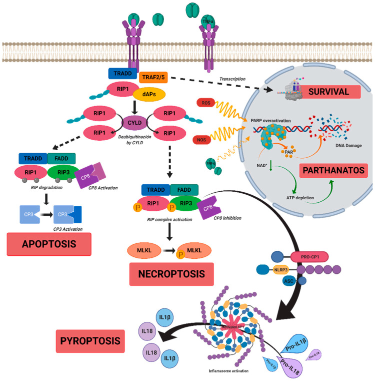
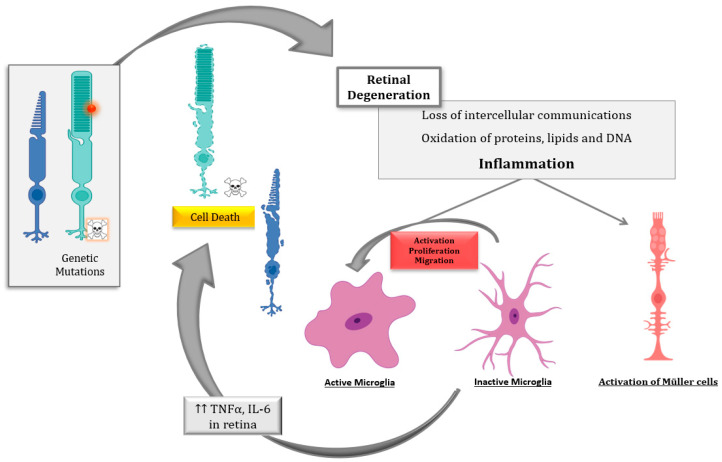
Inherited retinal dystrophies (IRDs) are a group of retinal disorders that cause progressive and severe loss of vision because of retinal cell death, mainly photoreceptor cells. IRDs include retinitis pigmentosa (RP), the most common IRD. IRDs present a genetic and clinical heterogeneity that makes it difficult to achieve proper treatment. The progression of IRDs is influenced, among other factors, by the activation of the immune cells (microglia, macrophages, etc.) and the release of inflammatory molecules such as chemokines and cytokines. Upregulation of tumor necrosis factor alpha (TNFα), a pro-inflammatory cytokine, is found in IRDs. This cytokine may influence photoreceptor cell death. Different cell death mechanisms are proposed, including apoptosis, necroptosis, pyroptosis, autophagy, excessive activation of calpains, or parthanatos for photoreceptor cell death. Some of these cell death mechanisms are linked to TNFα upregulation and inflammation. Therapeutic approaches that reduce retinal inflammation have emerged as useful therapies for slowing down the progression of IRDs. We focused this review on the relationship between retinal inflammation and the different cell death mechanisms involved in RP. We also reviewed the main anti-inflammatory therapies for the treatment of IRDs.

Keywords: TNFα; cell death; inflammation; retinal dystrophies.


Conflict of interest statement

The authors declare no conflict of interest. The funders had no role in the design of the study; in the collection, analyses, or interpretation of data; in the writing of the manuscript; or in the decision to publish the results.

Figures

Figure 1
Figure 1
A scheme of possible cell death mechanisms during of tumor necrosis factor alpha (TNFα)-induced signaling in inherited retinal dystrophies (IRDs). TNFα can simultaneously activate multiple signaling pathways of cell death or survival. TNFα binds to tumor necrosis factor receptor 1 (TNFR1), triggering three functional states. (i) The intracellular domain of TNFR1 recruits a death-domain containing adaptor protein (TRADD). TRADD recruits TNF receptor-associated factor 2 (TRAF2) and receptor-interacting protein kinase 1 (RIPK1) to form Complex 1. Complex 1 seems to be important for nuclear factor kappa beta (NF-κB)activation. NF-κB regulates anti-apoptotic genes to block the initiation of apoptosis by Complex 2. (ii) Complex 1 dissociates from TNFR1 and integrates Fas-associated protein with death domain (FADD) and pro-caspase 8 to form Complex 2. The FADD/caspase 8 association depends on complexes containing unubiquitinated RIPK1 as a scaffold. Activated caspase 8 induces caspase 3 and apoptosis. Under apoptotic conditions, active caspase 8 prevents further necroptotic signaling by cleaving and inactivating RIPK1 and RIPK3. (iii) Necroptosis is also mediated through TNFα signaling, when caspase 8 is not active. RIPK1 recruits RIPK3 to form the necrosome complex. RIPK3 phosphorylates the pseudokinase kinase-like domain of mixed-lineage kinase do-main-like (MLKL), leading to its oligomerization. Thus, MLKL recruitment to the plasma membrane induces necroptosis by triggering Ca+ and Na2+ influx into the cell. RIPK3 can also promote the nucleotide-binding oligomerization domain containing protein (NOD)-like receptor (NLR) family protein 3 (NLRP3) inflammasome and interleukin (IL)-1β inflammatory responses. TNFα or oxidative stress could activate parthanatos through the overactivation of poly [ADP-ribose] polymerase 1 (PARP1). PARP1 cleaves nicotinamide adenine dinucleotide (NAD+) to nicotinamide and adenosine diphosphate (ADP-ribose). PARPs couple one or more ADP-ribose (PAR) to acceptor proteins (PARylation) and components of the DNA repair machinery. Overactivation of PARP1 can deplete cellular adenosine triphosphate (ATP) and NAD+ storage and lead to bioenergetic collapse and cell death. Adapted from Olivares-González et al. [28].
Figure 2
Figure 2
Schematic representation of the inflammatory processes underlying photoreceptor degeneration in retinitis pigmentosa, the most common inherited retinal dystrophy (IRD). The genetic defect leads to rod degeneration. During rod degeneration several cellular processes are activated, including inflammation and oxidative stress, which lead to the loss of intercellular communications, the oxidation of macromolecules, and the activation of microglia and Müller cells, among others. Activated microglia cells release inflammatory molecules such as cytokines (tumor necrosis factor alpha (TNFα) and interleukin 6 (IL6)), chemokines, etc., which would exacerbate photoreceptor degeneration (both rods and cones) through different cell death mechanism such as necroptosis, parthanatos, apoptosis, etc.

References

    1. Hartong D.T., Berson E.L., Dryja T.P. Retinitis pigmentosa. Lancet. 2006;368:1795–1809. doi: 10.1016/S0140-6736(06)69740-7. - DOI - PubMed
    1. Wooff Y., Man S.M., Aggio-Bruce R., Natoli R., Fernando N. IL-1 Family Members Mediate Cell Death, Inflammation and Angiogenesis in Retinal Degenerative Diseases. Front. Immunol. 2019;10:1618. doi: 10.3389/fimmu.2019.01618. - DOI - PMC - PubMed
    1. Broadgate S., Yu J., Downes S.M., Halford S. Unravelling the genetics of inherited retinal dystrophies: Past, present and future. Prog. Retin. Eye Res. 2017;59:53–96. doi: 10.1016/j.preteyeres.2017.03.003. - DOI - PubMed
    1. RetNet Retinal Information Network. [(accessed on 29 December 2020)]; Available online: https://sph.uth.edu/retnet/home.htm.
    1. Chang B. Animal Models of Retinitis Pigmentosa (RP) In: Chan C.-C., editor. Animal Models of Ophthalmic Diseases. Springer International Publishing; Cham, Switzerland: 2016. pp. 101–116.

LinkOut - more resources