Review
. 2021 Feb 20;13(4):890.
doi: 10.3390/cancers13040890.

Targeting SHIP1 and SHIP2 in Cancer

Affiliations
Review

Targeting SHIP1 and SHIP2 in Cancer

Chiara Pedicone et al. Cancers (Basel). .

Abstract

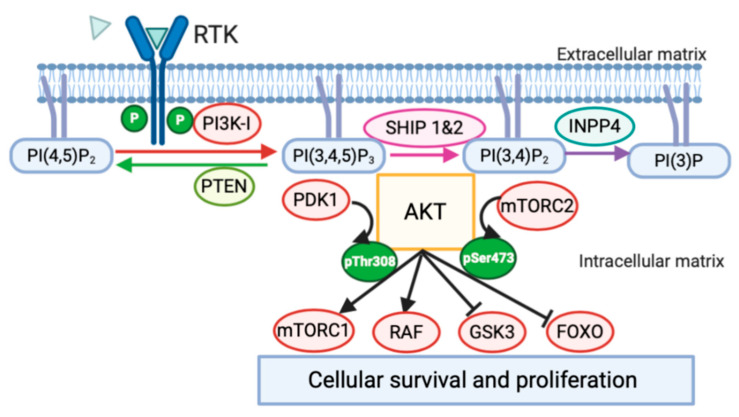
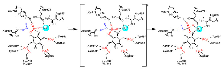
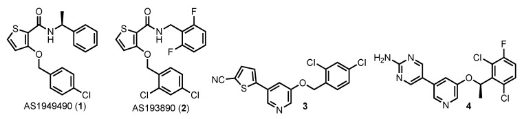
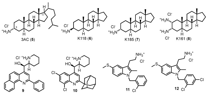
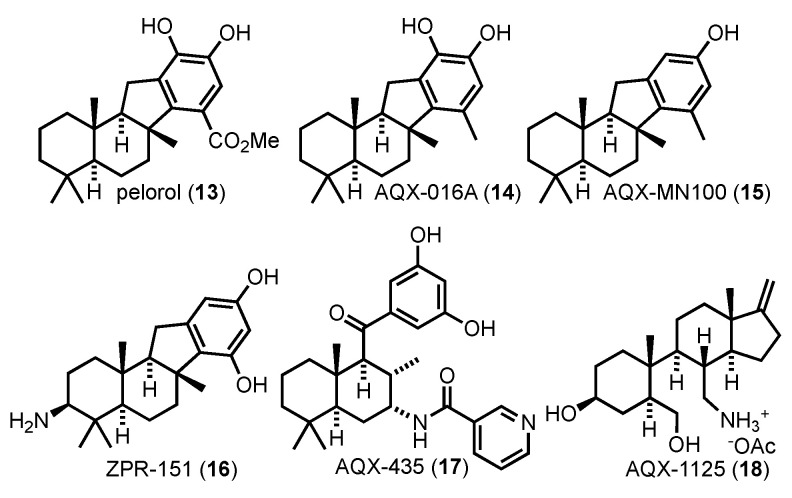
Membrane-anchored and soluble inositol phospholipid species are critical mediators of intracellular cell signaling cascades. Alterations in their normal production or degradation are implicated in the pathology of a number of disorders including cancer and pro-inflammatory conditions. The SH2-containing 5' inositol phosphatases, SHIP1 and SHIP2, play a fundamental role in these processes by depleting PI(3,4,5)P3, but also by producing PI(3,4)P2 at the inner leaflet of the plasma membrane. With the intent of targeting SHIP1 or SHIP2 selectively, or both paralogs simultaneously, small molecule inhibitors and agonists have been developed and tested in vitro and in vivo over the last decade in various disease models. These studies have shown promising results in various pre-clinical models of disease including cancer and tumor immunotherapy. In this review the potential use of SHIP inhibitors in cancer is discussed with particular attention to the molecular structure, binding site and efficacy of these SHIP inhibitors.

Keywords: AKT; Caspase 8; Fas; PI(3,4)P2; PI(3,4,5)P3; PI3K; SHIP1; SHIP2; SHIPi; cancer.


Conflict of interest statement

W.G.K., C.P. and J.D.C. have patents on small molecules targeting of SHIP1 and SHIP2 in disease. W.G.K. is Chief Scientific Officer and J.D.C. serves on the Scientific Advisory Board of the company “Alterna Therapeutics” for development and commercialization of SHIP inhibitor therapeutics. The other authors have no conflicts to disclose.

Figures

Figure 1
Figure 1
Modification of PtdIns Mediated by PI3K, PTEN, SHIP, INPP4 and the PI3K signaling pathway in cancer: The phosphorylated state of inositol lipids inserted in the cell membrane is controlled by inositol kinase and phosphatase enzymes. As proposed in “The Two PIP hypothesis” [22], the presence of both PI(3,4,5)P3 and PI(3,4)P2 at the membrane triggers downstream signaling events that more effectively promote cancer cell survival. Downstream targets of AKT are both pro-survival (activation of mTORC1, RAF) and anti-apoptotic (inhibition of GSK-3, FOXO) permitting increased cancer cell survival and proliferation.
Figure 2
Figure 2
SHIP protein structure and active sites SHIP1 and SHIP2 are multidomain proteins constituted of: SH2 domain, PH-L domain, 5′-phosphatase catalytic domain, C2 domain and C-terminal domain containing PXXP and NPXY motifs. (A) SHIP1 has phosphorylation sites on Serine 440 (Ser440) and Tyrosine 1020 (Tyr1020) that regulate respectively its activation and ubiquitination. (B) SHIP2 has a ubiquitin interacting motif (UIM) and a sterile alpha motif (SAM) domain, not present in SHIP1.
Figure 3
Figure 3
Overlap of X-ray structures of SHIP1 and SHIP2 containing C2 domain and phosphatase domain. (A) Structure of SHIP1 (in grey, 6IBD) and SHIP2 (in green, 5OKM) aligned with a model of PI(3,4,5)P3 constructed as described by Lietha [144], where coordinates for the inositol and metal ion are taken from PDB: 3MTC [146]. The C2 domain is on the left and the phosphatase domain is on the right. In the SHIP1 crystal the P4IM flexible loop region is in the “in” conformation (in blue), while in the SHIP2 crystal structure this portion of the enzyme is in the “out” position (in violet). (B) Closeup of the 5′-phosphate binding site from (A), SHIP1 in grey, SHIP2 in green. The residues in SHIP1 and SHIP2 have close homology in both structure and position. Some disorder is seen with Glu452 in the SHIP1 structure 6IBD, two conformations are given in the structure, but Glu473 in 5OKM overlaps with one of the conformers and likely can access the other with little difficulty.
Figure 4
Figure 4
Sequence comparison of the SHIP1 and SHIP2 P4IM region [146]. Conservative amino acid differences are highlighted in yellow, radical amino acid differences are highlighted in blue.
Figure 5
Figure 5
Catalysis of the cleavage of the 5′-phosphate of PI(3,4,5)P3 by SHIP2.
Figure 6
Figure 6
SHIP2 Inhibitors.
Figure 7
Figure 7
Aminosteroids, quinoline and tryptamine SHIP antagonists from Kerr and co-workers.
Figure 8
Figure 8
SHIP1 Agonists.

References

    1. Zhang X., Majerus P.W. Phosphatidylinositol signaling reactions. Semin. Cell Dev. Biol. 1998;9:153–160. doi: 10.1006/scdb.1997.0220. - DOI - PubMed
    1. Catimel B., Yin M.-X., Schieber C., Condron M., Patsiouras H., Catimel J., Robinson D.E.J.E., Wong L.S.-M., Nice E.C., Holmes A.B., et al. PI(3,4,5)P3 Interactome. J. Proteome Res. 2009;8:3712–3726. doi: 10.1021/pr900320a. - DOI - PubMed
    1. Falkenburger B.H., Jensen J.B., Dickson E.J., Suh B.-C., Hille B. Phosphoinositides: Lipid regulators of membrane proteins. J. Physiol. 2010;588:3179–3185. doi: 10.1113/jphysiol.2010.192153. - DOI - PMC - PubMed
    1. Franke T.F., Kaplan D.R., Cantley L.C., Toker A. Direct regulation of the Akt proto-oncogene product by phosphatidylinositol-3,4-bisphosphate. Science. 1997;275:665–668. doi: 10.1126/science.275.5300.665. - DOI - PubMed
    1. Ma K., Cheung S.M., Marshall A.J., Duronio V. PI(3,4,5)P3 and PI(3,4)P2 levels correlate with PKB/akt phosphorylation at Thr308 and Ser473, respectively; PI(3,4)P2 levels determine PKB activity. Cell Signal. 2008;20:684–694. doi: 10.1016/j.cellsig.2007.12.004. - DOI - PubMed

LinkOut - more resources