. 2021 Feb 14;11(2):278.
doi: 10.3390/biom11020278.

Valency and Binding Affinity Variations Can Regulate the Multilayered Organization of Protein Condensates with Many Components

Affiliations

Valency and Binding Affinity Variations Can Regulate the Multilayered Organization of Protein Condensates with Many Components

Ignacio Sanchez-Burgos et al. Biomolecules. .

Abstract

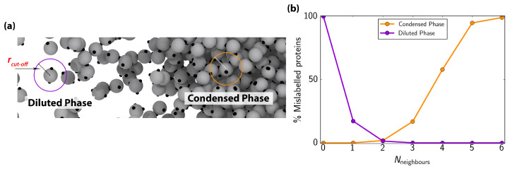
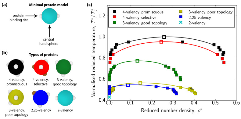
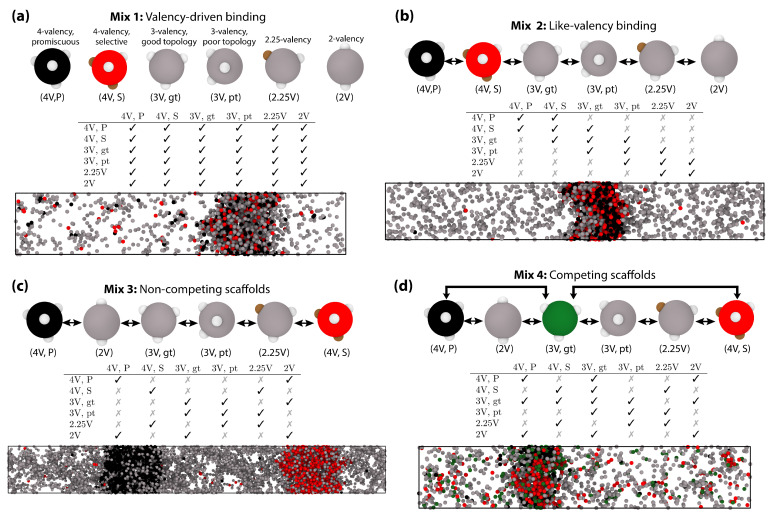
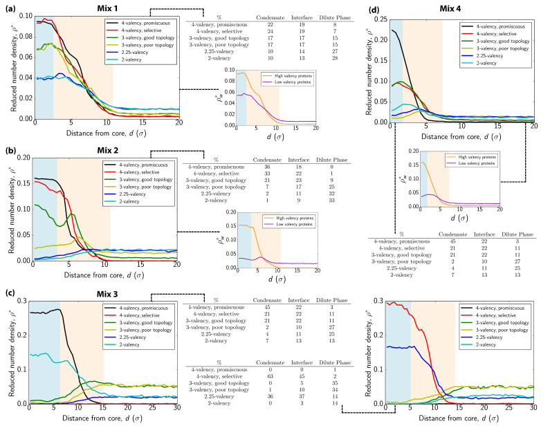
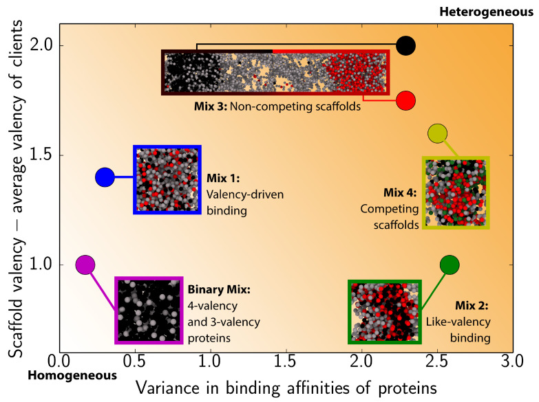
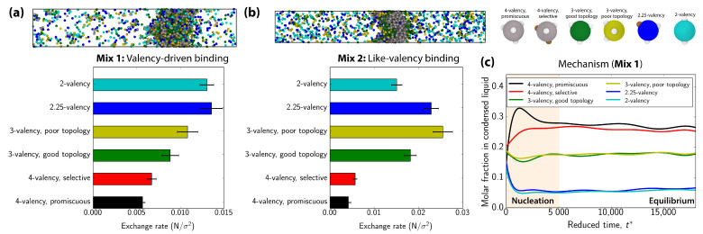
Biomolecular condensates, which assemble via the process of liquid-liquid phase separation (LLPS), are multicomponent compartments found ubiquitously inside cells. Experiments and simulations have shown that biomolecular condensates with many components can exhibit multilayered organizations. Using a minimal coarse-grained model for interacting multivalent proteins, we investigate the thermodynamic parameters governing the formation of multilayered condensates through changes in protein valency and binding affinity. We focus on multicomponent condensates formed by scaffold proteins (high-valency proteins that can phase separate on their own via homotypic interactions) and clients (proteins recruited to condensates via heterotypic scaffold-client interactions). We demonstrate that higher valency species are sequestered to the center of the multicomponent condensates, while lower valency proteins cluster towards the condensate interface. Such multilayered condensate architecture maximizes the density of LLPS-stabilizing molecular interactions, while simultaneously reducing the surface tension of the condensates. In addition, multilayered condensates exhibit rapid exchanges of low valency proteins in and out, while keeping higher valency proteins-the key biomolecules involved in condensate nucleation-mostly within. We also demonstrate how modulating the binding affinities among the different proteins in a multicomponent condensate can significantly transform its multilayered structure, and even trigger fission of a condensate into multiple droplets with different compositions.

Keywords: minimal protein model; multicomponent condensates; multilayered condensates; multiphase condensates; protein liquid–liquid phase separation.


Conflict of interest statement

The authors declare no conflict of interest. The funders had no role in the design of the study; in the collection, analyses, or interpretation of data; in the writing of the manuscript, or in the decision to publish the results.

Figures

Figure A1
Figure A1
(a) Snapshot of the interfacial region between the two coexisting phases of a 6-component mixture. The order parameter distinguishes between proteins (in grey) of both phases by evaluating the number of neighbours that each protein has for a given cut-off distance. For considering a protein belonging to the condensed phase, it needs to have at least 2 neighbours (independent of its valency) within a cut-off distance of rcut-off=1.20σ. (b) Percentage of mislabelled proteins as a function of the number of neighbours in the diluted (purple curve) and in the condensed phase (orange curve) for a cut-off distance of 1.20σ. Both curves (phases) were evaluated in bulk NVT simulations at the coexisting density and composition of the ‘valency-driven binding’ system at T* = 0.09.
Figure 1
Figure 1
(a) Minimal protein model. In our coarse-grained model, proteins are represented by hard-spheres with different numbers/types of binding sites on their surface. (b) Schematic representation of the different multivalent proteins studied in this work. 4-valency promiscuous proteins have four promiscuous binding sites in a tetrahedral arrangement. 4-valency selective proteins have two types of binding sites that interact exclusively with those of the same type on their parent protein (i.e., selective homotypic binding), and promiscuously with any site on their non-parent proteins. In total, this protein has four patches arranged in a tetrahedral topology. 3-valency good topology proteins have three promiscuous binding sites separated by angles of 120 in a plane, which minimizes the steric hindrance for binding. 3-valency ‘poor’ topology proteins have binding sites separated by 90 angles, which leads to larger steric hindrance. 2.25-valency proteins possess the same topology as the 3-valency good topology proteins, but the strength of one of the binding sites (brown) is decreased to 1/4 of the net strength interaction of the other two sites (i.e., brown–brown = 25% white–white). Interactions between that special site and regular sites follow Lorentz-Berthelot combination rule (ϵ1+ϵ22). 2-valency proteins have two binding sites in a polar arrangement. These snapshots and subsequent ones were rendered using OVITO [85]. (c) Phase diagrams in the (T*/Tc*) ρ* plane for the 6 different proteins considered in this study. T*/Tc* is the reduced temperature normalized by the highest critical reduced temperature [corresponding to the 4-valency promiscuous protein (Tc* = 0.121)] and ρ* is the reduced density. Filled squares represent the coexistence points computed using Direct Coexistence simulations. Empty squares depict the estimated critical points using the universal scaling of the coexistence densities near the critical point, and the law of rectilinear diameters [86]. Further details on the reduced units used in this work are given in the Appendix A. Note that the 2-valency protein does not phase separate on its own (cyan cross).
Figure 2
Figure 2
(a) Valency-driven binding. In this system, all interactions between different protein types are enabled; the table inset describes which proteins bind to one another with high-affinity (tick) or low-affinity (cross). A representative snapshot of the coexisting condensate with the dilute phase at T*=0.09 taken from a Direct Coexistence (DC) simulation is shown. Here, 4-valency promiscuous binding proteins and 4-valency selective binding ones are colored in black and red respectively, while lower valency proteins are colored in grey (as shown in the snapshots, top panel). (b) Like-valency binding. In this system, proteins participate in either homotypic binding or interact with other proteins of like-valency, as indicated in the table inset. A snapshot of this system at T*=0.09 is shown, with the same color code as in (a). (c) Non-competing scaffolds. Homotypic interactions are allowed (see diagonal of the table inset) plus the heterotypic interactions indicated in the table. In this case, the two highest valency proteins (i.e., 4-valency promiscuous and 4-valency selective) cannot bind to each other. For this interaction scheme, two different condensates with different compositions are formed in our DC simulations at T*=0.083. Note that for this system, no phase separation was observed at T*=0.09. (d) Competing scaffolds. Similar binding scheme as in (c), except that the 2 highest valency proteins now compete for binding to the 3-valency good topology protein (see table inset). In this case, the system condenses into a single droplet, despite the absence of attractive interactions between 4-valency promiscuous proteins and 4-valency selective ones. A representative snapshot of a DC simulation at T*=0.09 is shown. The same color code as in a–c is used, except for the 3-valency good topology proteins, which are now depicted in green.
Figure 3
Figure 3
Structural insights of multilayer and multidroplet organization. (ad) Density profiles of each protein type for the systems depicted in Figure 2a–d, respectively. In each profile, we plot the reduced density (ρ*) of a given protein as a function of distance (in units of molecular diameter, σ) from the droplet center of mass along the perpendicular direction to the interface (long axis of the simulation box). The protein concentration of each system in the condensate (blue shaded region), interfacial boundaries (beige shaded region) and dilute phase (white region) are reported for each mixture. Density profiles of the different mixtures in terms of high-valency proteins (4-valency) vs low-valency ones (3- and 2-valency) are also given for mixtures (a,b,d). Since the number of low-valency species is double than that of high-valency ones by construction, we plot weighted density ρw* (total density of scaffolds/clients divided by the number of protein types belonging to each family) against distance from the center of mass of the droplet. Note that for mixture (c) we display two density profiles corresponding to each of the droplets observed in Figure 2c: (right) the 4-valency promiscuous rich one and (left) the 4-valency selective rich droplet. The temperatures at which these analyses were performed are those indicated in Figure 2.
Figure 4
Figure 4
Relationship between valency and binding affinity for the different studied mixtures. On the y-axis, we plot the difference between the scaffold valency and the average valency of clients. Here, scaffolds are: 4-valency promiscuous proteins for the Binary Mix [41] and for the black condensate in Mix 3; both 4-valency proteins (i.e., promiscuous and selective) in Mix 1, 2, and 4; and 4-valency selective proteins for the red condensate in Mix 3. Note, we consider separate scaffolds for Mix 3; since, in that system, the two types of 4-valency proteins form distinct condensates. To calculate the average valency of clients, we only consider clients that can bind to the respective scaffolds with a high binding affinity (i.e., non-zero in this case; Figure 2). The x-axis represents the variance in pairwise binding affinities for all the proteins in the system; where the binding affinity for a given pairwise interaction is taken as the average valency of the two proteins in question (see table insets in Figure 2 for details on pairwise binding interactions). An increase in either the scaffold–client valency difference or the variance in protein binding affinities lead to progressively more heterogeneous condensates, as indicated by the orange shaded background.
Figure 5
Figure 5
(a,b) Exchange rate between the condensate and the dilute phase for proteins in systems (a) and (b) shown in Figure 2 and Figure 3. The exchange rate is defined as the averaged difference in the number of proteins per unit of area (N/σ2; in our calculations, the interfacial area of the slab is two times the cross-section of our DC simulation box) for each component between subsequent independent configurations. One configuration is considered independent from the previous one when the proteins inside the condensate have diffused at least one molecular diameter. For both mixtures, a snapshot of the direct coexistence simulation from which exchange rate calculations were performed is shown. 4-valency proteins are colored in grey. The coloring code for all other proteins is the same as that used in the exchange rate diagrams. (c) Time-evolution of the molar fraction of each individual specie in the condensed liquid phase as a function of time (in reduced units) for the valency-driven system (Mix 1). t* = 0 corresponds to the homogeneous fluid state of the 6-component mixture. The temperature of all the NVT simulations shown in this Figure is T* = 0.09.

Similar articles

Cited by

References

    1. Sear R.P. The cytoplasm of living cells: A functional mixture of thousands of components. J. Phys. Condens. Matter. 2005;17:S3587–S3595. doi: 10.1088/0953-8984/17/45/052. - DOI
    1. Hyman A.A., Weber C.A., Jülicher F. Liquid-Liquid Phase Separation in Biology. Annu. Rev. Cell Dev. Biol. 2014;30:39–58. doi: 10.1146/annurev-cellbio-100913-013325. - DOI - PubMed
    1. Banani S.F., Lee H.O., Hyman A.A., Rosen M.K. Biomolecular condensates: Organizers of cellular biochemistry. Nat. Rev. Mol. Cell Biol. 2017;18:285–298. doi: 10.1038/nrm.2017.7. - DOI - PMC - PubMed
    1. Shin Y., Brangwynne C.P. Liquid phase condensation in cell physiology and disease. Science. 2017;357:eaaf4382. doi: 10.1126/science.aaf4382. - DOI - PubMed
    1. Su X., Ditlev J.A., Hui E., Xing W., Banjade S., Okrut J., King D.S., Taunton J., Rosen M.K., Vale R.D. Phase separation of signaling molecules promotes T cell receptor signal transduction. Science. 2016;352:595–599. doi: 10.1126/science.aad9964. - DOI - PMC - PubMed

LinkOut - more resources