Review
. 2021 Feb 14;13(4):790.
doi: 10.3390/cancers13040790.

Performance of Ultrasound Techniques and the Potential of Artificial Intelligence in the Evaluation of Hepatocellular Carcinoma and Non-Alcoholic Fatty Liver Disease

Affiliations
Review

Performance of Ultrasound Techniques and the Potential of Artificial Intelligence in the Evaluation of Hepatocellular Carcinoma and Non-Alcoholic Fatty Liver Disease

Monica Lupsor-Platon et al. Cancers (Basel). .

Abstract

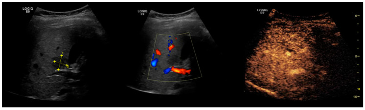
Global statistics show an increasing percentage of patients that develop non-alcoholic fatty liver disease (NAFLD) and NAFLD-related hepatocellular carcinoma (HCC), even in the absence of cirrhosis. In the present review, we analyzed the diagnostic performance of ultrasonography (US) in the non-invasive evaluation of NAFLD and NAFLD-related HCC, as well as possibilities of optimizing US diagnosis with the help of artificial intelligence (AI) assistance. To date, US is the first-line examination recommended in the screening of patients with clinical suspicion of NAFLD, as it is readily available and leads to a better disease-specific surveillance. However, the conventional US presents limitations that significantly hamper its applicability in quantifying NAFLD and accurately characterizing a given focal liver lesion (FLL). Ultrasound contrast agents (UCAs) are an essential add-on to the conventional B-mode US and to the Doppler US that further empower this method, allowing the evaluation of the enhancement properties and the vascular architecture of FLLs, in comparison to the background parenchyma. The current paper also explores the new universe of AI and the various implications of deep learning algorithms in the evaluation of NAFLD and NAFLD-related HCC through US methods, concluding that it could potentially be a game changer for patient care.

Keywords: artificial intelligence; contrast enhanced ultrasound; focal liver lesion; hepatocellular carcinoma; non-alcoholic fatty liver disease; steatosis; ultrasonography.


Conflict of interest statement

The authors declare no conflict of interest.

Figures

Figure 1
Figure 1
Hepatic steatosis. The 2D-US examination is showing an enlarged liver, with increased echogenicity and posterior beam attenuation, with a slightly inhomogeneous structure of fine granularity, without any FLLs. Even if this aspect is highly suggestive of hepatic steatosis, conventional US is unable to properly quantify the fatty amount of the liver. Also, 2D-US cannot specify whether fibrosis is present or not. Usually, steatosis and fibrosis coexist and therefore “steato-fibrosis” is the preferred term in this situation.
Figure 2
Figure 2
Hepatic steatosis. Focal liver lesion. 2D-US scan shows an enlarged liver with increased echogenicity and posterior beam attenuation. In addition, a focal parenchymal structure is observed in the right lobe. It is characterized as having decreased, heterogeneous echogenicity and internal vessels seen at Doppler examination. The diagnosis of the focal lesion remains uncertain and CEUS examination is necessary.
Figure 3
Figure 3
Hepatic steatosis. HCC. Conventional B-mode image (a), CEUS in the arterial phase (b), and CEUS in the late phase (c). The examination shows an enlarged liver with markedly increased echogenicity and a focal parenchymal lesion with decreased echogenicity (a). At CEUS technique, the lesion shows enhancement in the arterial phase (b), followed by washout in the late phase (c).
Figure 4
Figure 4
CEUS LI-RADS classification and management of FLLs according to the American College of Radiology [59,111]. This table includes only CEUS pure blood pool agents (SonoVue, Luminity). * MDD: multidisciplinary discussion; #MDD should be considered, since a recent prospective study found that 60% of CEUS LR-3 observations were HCCs [112].
Figure 5
Figure 5
Focal sparing in the pericholecystic region. Hypoechoic image located around the gallbladder in a fatty liver. The Doppler examination cannot reveal any vessels within the lesion. On CEUS, the enhancement was homogeneous during the arterial, portal, and parenchymal phases, without any apparent focal lesions.

Similar articles

Cited by

References

    1. Estes C., Razavi H., Loomba R., Younossi Z., Sanyal A.J. Modeling the epidemic of nonalcoholic fatty liver disease demonstrates an exponential increase in burden of disease. Hepatology. 2018;67:123–133. doi: 10.1002/hep.29466. - DOI - PMC - PubMed
    1. Younossi Z.M. Non-alcoholic fatty liver disease—A global public health perspective. J. Hepatol. 2019;70:531–544. doi: 10.1016/j.jhep.2018.10.033. - DOI - PubMed
    1. Singh S., Muir A.J., Dieterich D.T., Falck-Ytter Y.T. American Gastroenterological Association Institute Technical Review on the Role of Elastography in Chronic Liver Diseases. Gastroenterology. 2017;152:1544–1577. doi: 10.1053/j.gastro.2017.03.016. - DOI - PubMed
    1. Maher J.J., Schattenberg J.M. Nonalcoholic Fatty Liver Disease in 2020. Gastroenterology. 2020;158:1849–1850. doi: 10.1053/j.gastro.2020.04.013. - DOI - PMC - PubMed
    1. Papatheodoridi M., Cholongitas E. Diagnosis of Non-alcoholic Fatty Liver Disease (NAFLD): Current Concepts. Curr. Pharm. Des. 2018;24:4574–4586. doi: 10.2174/1381612825666190117102111. - DOI - PubMed

LinkOut - more resources