

Emerging Utility of Applied Magnetic Resonance Imaging in the Management of Traumatic Brain Injury
- PMID: 33673012
- PMCID: PMC7930990
- DOI: 10.3390/medsci9010010
Emerging Utility of Applied Magnetic Resonance Imaging in the Management of Traumatic Brain Injury
Abstract
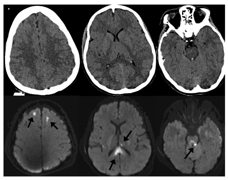
Traumatic brain injury (TBI) is a widespread and expensive problem globally. The standard diagnostic workup for new TBI includes obtaining a noncontrast computed tomography image of the head, which provides quick information on operative pathologies. However, given the limited sensitivity of computed tomography for identifying subtle but meaningful changes in the brain, magnetic resonance imaging (MRI) has shown better utility for ongoing management and prognostication after TBI. In recent years, advanced applications of MRI have been further studied and are being implemented as clinical tools to help guide care. These include functional MRI, diffusion tensor imaging, MR perfusion, and MR spectroscopy. In this review, we discuss the scientific basis of each of the above techniques, the literature supporting their use in TBI, and how they may be clinically implemented to improve the care of TBI patients.
Keywords: MR perfusion; MR spectroscopy; diffusion tensor imaging; functional MRI; magnetic resonance imaging; traumatic brain injury.
Conflict of interest statement
The authors declare no conflict of interest.
Figures
References
-
- James S.L., Theadom A., Ellenbogen R.G., Bannick M.S., Montjoy-Venning W., Lucchesi L.R., Abbasi N., Abdulkader R., Abraha H.N., Adsuar J.C., et al. Global, regional, and national burden of traumatic brain injury and spinal cord injury, 1990–2016: A systematic analysis for the Global Burden of Disease Study 2016. Lancet Neurol. 2019;18:56–87. doi: 10.1016/S1474-4422(18)30415-0. - DOI - PMC - PubMed
-
- Vos T., Abajobir A.A., Abate K.H., Abbafati C., Abbas K.M., Abd-Allah F., Abdulkader R.S., Abdulle A.M., Abebo T.A., Abera S.F., et al. Global, regional, and national incidence, prevalence, and years lived with disability for 328 diseases and injuries for 195 countries, 1990–2016: A systematic analysis for the Global Burden of Disease Study 2016. Lancet. 2017;390:1211–1259. doi: 10.1016/S0140-6736(17)32154-2. - DOI - PMC - PubMed
Publication types
MeSH terms
LinkOut - more resources
Full Text Sources
Other Literature Sources
Medical
