Review
. 2021 Feb 12;22(4):1821.
doi: 10.3390/ijms22041821.

Phenotypic Plasticity of Cancer Cells Based on Remodeling of the Actin Cytoskeleton and Adhesive Structures

Affiliations
Review

Phenotypic Plasticity of Cancer Cells Based on Remodeling of the Actin Cytoskeleton and Adhesive Structures

Svetlana N Rubtsova et al. Int J Mol Sci. .

Abstract

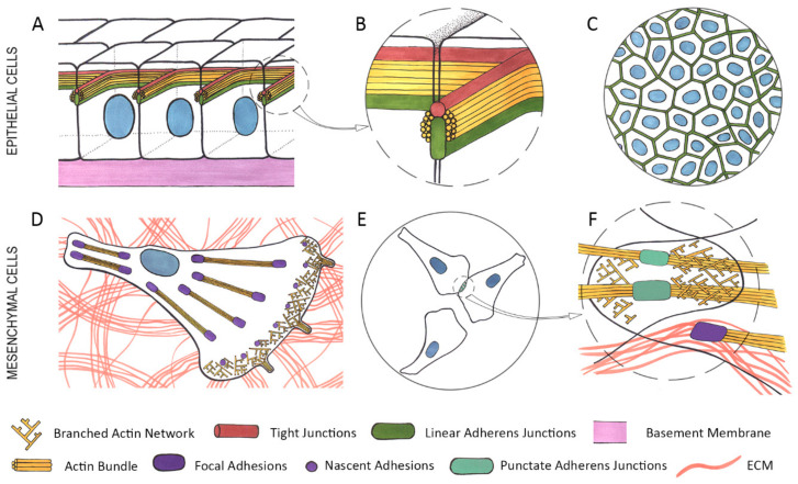
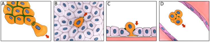
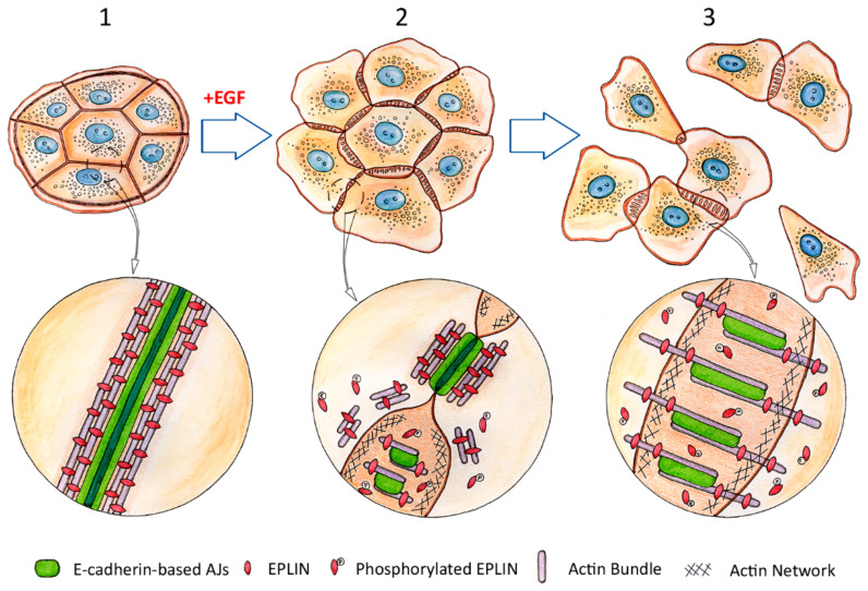
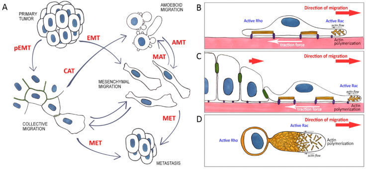
There is ample evidence that, instead of a binary switch, epithelial-mesenchymal transition (EMT) in cancer results in a flexible array of phenotypes, each one uniquely suited to a stage in the invasion-metastasis cascade. The phenotypic plasticity of epithelium-derived cancer cells gives them an edge in surviving and thriving in alien environments. This review describes in detail the actin cytoskeleton and E-cadherin-based adherens junction rearrangements that cancer cells need to implement in order to achieve the advantageous epithelial/mesenchymal phenotype and plasticity of migratory phenotypes that can arise from partial EMT.

Keywords: E-cadherin; EMT; actin cytoskeleton; adherens junctions; cancer cells; migration; plasticity.


Conflict of interest statement

The authors declare no conflict of interest.

Figures

Figure 1
Figure 1
Organization of the actin cytoskeleton and adhesive structures in epithelial and mesenchymal cells. (A)—a monolayer of epithelial cells. (B)—a close-up of an area in the dashed circle on A—stable cell-cell adhesion in epithelial cells is provided by apical adhesion belts comprised by tight junctions (TJs) (red) and linear adherens junctions (AJs) (green), both of which are closely associated with the circumferential actin bundle (yellow). (C)—a top view of a monolayer of epithelial cells, connected by stable linear AJs. (D)—a mesenchymal cell exhibiting branched actin network (yellow) and nascent focal adhesions (FAs) (purple) in lamellipodia at the leading edge. Closer to the center of the cell and in the rear are mature FAs (purple) associated with straight actin bindles (yellow). Both nascent and mature FAs are connected to the extracellular matrix (ECM) (pink). (E)—an area of cell-cell interaction between motile mesenchymal cells. (F)—a close-up of the area in the dashed circle on E—overlapping lamellae containing branched actin network (yellow) point to the lack of contact paralysis, unstable punctate AJs (green) are associated with straight actin bundles. Mature FAs (purple) connected to the ECM (pink) are associated with straight actin bundles.
Figure 2
Figure 2
Structure and composition of an epithelial adherens junction. E-cadherin molecules (green) on the surface of adjacent cells connect with one another via their extracellular domains. Below the cytoplasmic membrane (PM), the intracellular domain of E-cadherin interacts with p120 (pink) and β-catenins (dull red). β-catenin binds to α-catenin (dark blue), which, in turn, interacts with vinculin (blue) and directly with actin filaments (yellow). Vinculin binds to actin filaments to stabilize the cadherin-catenin complex. Various actin-binding proteins such as VASP (bright red), EPLIN (Epithelial Protein Lost in Neoplasm) (purple), α-actinin (orange), palladin (light green) and myosin II (cyan-green) are associated with junctional actin.
Figure 3
Figure 3
E-cadherin-based adherens junctions (AJs) facilitate cancer cell dissemination. (A)—collective invasion. (B)—migration over a monolayer of normal epithelial cells. (C)—invasion of the monolayer of normal epithelial cells. (D)—a circulating tumor cell (CTC) cluster traveling through circulation. Orange—cancer cells, grey—normal epithelial cells, purple—endothelial cells. Green—E-cadherin-based AJs, red arrows—direction of cell migration.
Figure 4
Figure 4
Dissolution of the circumferential actin bundle, degradation of EPLIN and rearrangement of E-cadherin-based adherens junctions (AJs) during the early stages of epithelial-mesenchymal transition (EMT). Adapted from Zhitnyak et al., Cells 2020, 9, 578 [150]. Top row: progression of EMT induced by Epidermal Growth Factor (EGF). 1—epithelial cells before treatment with EGF. An island of non-motile cells tightly connected by linear AJs. Circled area in higher magnification below: a stable linear AJ between epithelial cells, associated with a robust circumferential actin bundle. EPLIN supports the bundle integrity by cross-linking actin filaments. 2—early stages of EGF-induced EMT (5–10 min). Protrusive activity increases at the cell edges and protrusions begin to form at the cell-cell boundaries. Contact paralysis at the cell-cell boundaries disappears. Linear AJs are partially disassembled and replaced by punctate AJs. Circled area in higher magnification below: Dissolution of the circumferential actin bundle and reorganization of linear AJs. Part of the AJ still maintains its original linear configuration and is associated with the remnants of the circumferential actin bundle. Phosphorylation of EPLIN results in its detachment from the circumferential actin bundle which leads to disintegration of the bundle. Bottom part—formation of small punctate AJs associated with nascent straight actin bundles. 3—later stages of EGF-induced EMT (15–60 min). Cells acquire migratory properties and detach from each other, breaking cell-cell adhesion. The new punctate AJs formed by migratory cells are dynamic and unstable. Circled area in higher magnification below: mature punctate AJs during later stages of EMT. The AJs are longer than in 2 and are associated with thicker straight actin bundles again fortified by EPLIN.
Figure 5
Figure 5
Plasticity of cancer cell migration. (A)—A primary epithelial tumor through partial epithelial- mesenchymal transition (pEMT) or epithelial-mesenchymal transtion (EMT) gives rise to motile cells capable of invasion via individual or collective mesenchymal migration. Specific microenvironment conditions govern reversible transitions between mesenchymal and amoeboid migration modes (collective-amoeboid transition (CAT), mesenchymal-amoeboid transition (MAT), amoeboid-mesenchymal transition (AMT)). In a distant metastasis, cells cease to migrate and revert to the original epithelial phenotype (mesenchymal-epithelial transition (MET)). (BD)—Modes of cancer cell migration. Reorganized actin cytoskeleton and relative activity of Rho and Rac in the front and rear of the cells during individual (B), collective (C), or amoeboid (D) migration.

Similar articles

Cited by

References

    1. Takeichi M. Dynamic contacts: Rearranging adherens junctions to drive epithelial remodelling. Nat. Rev. Mol. Cell. Biol. 2014;15:397–410. doi: 10.1038/nrm3802. - DOI - PubMed
    1. Vasileva E., Citi S. The role of microtubules in the regulation of epithelial junctions. Tissue Barriers. 2018;6:1539596. doi: 10.1080/21688370.2018.1539596. - DOI - PMC - PubMed
    1. Riga A., Castiglioni V.G., Boxem M. New insights into apical-basal polarization in epithelia. Curr. Opin. Cell Biol. 2020;62:1–8. doi: 10.1016/j.ceb.2019.07.017. - DOI - PubMed
    1. Takeichi M. Self-organization of animal tissues: Cadherin-mediated processes. Dev. Cell. 2011;21:24–26. doi: 10.1016/j.devcel.2011.06.002. - DOI - PubMed
    1. Davis M.A., Ireton R.C., Reynolds A.B. A core function for p120-catenin in cadherin turnover. J. Cell Biol. 2003;163:525–534. doi: 10.1083/jcb.200307111. - DOI - PMC - PubMed

MeSH terms