Review
. 2021 Feb 12;9(2):185.
doi: 10.3390/biomedicines9020185.

Evaluation of Plaque Characteristics and Inflammation Using Magnetic Resonance Imaging

Affiliations
Review

Evaluation of Plaque Characteristics and Inflammation Using Magnetic Resonance Imaging

Kristina Andelovic et al. Biomedicines. .

Abstract

Atherosclerosis is an inflammatory disease of large and medium-sized arteries, characterized by the growth of atherosclerotic lesions (plaques). These plaques often develop at inner curvatures of arteries, branchpoints, and bifurcations, where the endothelial wall shear stress is low and oscillatory. In conjunction with other processes such as lipid deposition, biomechanical factors lead to local vascular inflammation and plaque growth. There is also evidence that low and oscillatory shear stress contribute to arterial remodeling, entailing a loss in arterial elasticity and, therefore, an increased pulse-wave velocity. Although altered shear stress profiles, elasticity and inflammation are closely intertwined and critical for plaque growth, preclinical and clinical investigations for atherosclerosis mostly focus on the investigation of one of these parameters only due to the experimental limitations. However, cardiovascular magnetic resonance imaging (MRI) has been demonstrated to be a potent tool which can be used to provide insights into a large range of biological parameters in one experimental session. It enables the evaluation of the dynamic process of atherosclerotic lesion formation without the need for harmful radiation. Flow-sensitive MRI provides the assessment of hemodynamic parameters such as wall shear stress and pulse wave velocity which may replace invasive and radiation-based techniques for imaging of the vascular function and the characterization of early plaque development. In combination with inflammation imaging, the analyses and correlations of these parameters could not only significantly advance basic preclinical investigations of atherosclerotic lesion formation and progression, but also the diagnostic clinical evaluation for early identification of high-risk plaques, which are prone to rupture. In this review, we summarize the key applications of magnetic resonance imaging for the evaluation of plaque characteristics through flow sensitive and morphological measurements. The simultaneous measurements of functional and structural parameters will further preclinical research on atherosclerosis and has the potential to fundamentally improve the detection of inflammation and vulnerable plaques in patients.

Keywords: arterial elasticity; atherosclerosis; inflammation; magnetic resonance imaging; mouse models; pulse wave velocity; wall shear stress.


Conflict of interest statement

The authors declare no conflict of interest.

Figures

Figure 1
Figure 1
Four-dimensional (4D) flow and wall shear stress (WSS) measurements in the murine aortic arch. (A) Wall shear stress map of the aortic arch of an ApoE−/− mouse fed a Western diet, assessed with a self-gated radial 4D phase-contrast magnetic resonance imaging (PC-MRI) sequence [79]. Especially in the ascending aortic arch, a large region of low WSS can be observed in the inner curvature. This area is known to be prone to plaque development. (B) Streamline presentation of the measured flow and through plane flow for 6 exemplary analysis planes, obtained from the same 4D flow measurement [79,80].
Figure 2
Figure 2
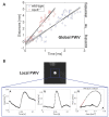
Global and local measurements of pulse wave velocity in murine vessels. (A) Assessment of global pulse wave velocity (PWV) for exemplary measurements in the aortic arch of a wild-type and an ApoE−/− mouse. For determination of the global PWV values, the time points of the early systolic upstrokes were measured for multiple planes along the aortic arch (see Figure 1B) and plotted against the locations of the planes. The PWV is derived from the slope of a line fitted to the data points [80]. (B) Exemplary measurement of the local PWV in the abdominal aorta of a wild-type mouse using a retrospectively navigated PC-Cine MRI technique [121]. The through-plane flow (Q) and the cross-sectional areas (A) were determined. The local PWV is derived from the Q-A curve by fitting a line to the early systolic data points.
Figure 3
Figure 3
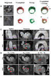
Morphology and inflammation imaging in murine atherosclerotic vessels. (A) Multispin multi echo (MSME) imaging of the ascending aorta (A) and the carotid artery (B) of an ApoE/ mouse fed a western diet. FG: Fibrotic tissue. L: Lipid rich core. C: Calcified tissue. (C,D) 3D VCAM-1 (vascular cell adhesion molecule-1) Measurement [141]. (C) Sagittal, transverse, and coronal orientation of the aorta extracted from the radial 3D-Cine measurement (TE1 = 1.5 ms) before administration of the contrast agent (pre) and after administration of the contrast agent (post). The lines mark the slice orientations. Areas of signal cancellation due to iron particles are marked with red arrows. (D) Measurement of the aortic arch at one exemplary position (see purple line in Figure 3C). Measurement with TE1 = 1.5 ms, TE2 = 2.3 ms and map of the phase differences Φ = TE2 − TE1. No significant phase jumps can be found in the aortic vessel wall in the pre-contrast agent measurement (red arrow, pre), whereas in the phase difference map of the post-contrast agent (CA) measurement (see red arrow in bottom right image (post)), significant phase jumps are observable.

References

    1. Libby P., Ridker P.M., Hansson G.K. Inflammation in atherosclerosis: From pathophysiology to practice. J. Am. Coll. Cardiol. 2009;54:2129–2138. doi: 10.1016/j.jacc.2009.09.009. - DOI - PMC - PubMed
    1. Bäck M., Yurdagul A., Tabas I., Öörni K., Kovanen P.T. Inflammation and its resolution in atherosclerosis: Mediators and therapeutic opportunities. Nat. Rev. Cardiol. 2019;16:389–406. doi: 10.1038/s41569-019-0169-2. - DOI - PMC - PubMed
    1. Wolf D., Ley K. Immunity and Inflammation in Atherosclerosis. Circ. Res. 2019;124:315–327. doi: 10.1161/CIRCRESAHA.118.313591. - DOI - PMC - PubMed
    1. Moriya J. Critical roles of inflammation in atherosclerosis. J. Cardiol. 2019;73:22–27. doi: 10.1016/j.jjcc.2018.05.010. - DOI - PubMed
    1. Ridker P.M., Everett B.M., Thuren T., MacFadyen J.G., Chang W.H., Ballantyne C., Fonseca F., Nicolau J., Koenig W., Anker S.D., et al. Antiinflammatory Therapy with Canakinumab for Atherosclerotic Disease. N. Engl. J. Med. 2017;377:1119–1131. doi: 10.1056/NEJMoa1707914. - DOI - PubMed

LinkOut - more resources