

Multi-Omic Biomarkers as Potential Tools for the Characterisation of Pancreatic Cystic Lesions and Cancer: Innovative Patient Data Integration
- PMID: 33673153
- PMCID: PMC7918773
- DOI: 10.3390/cancers13040769
Multi-Omic Biomarkers as Potential Tools for the Characterisation of Pancreatic Cystic Lesions and Cancer: Innovative Patient Data Integration
Abstract
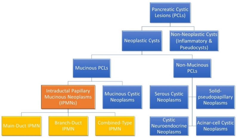
Pancreatic cancer (PC) is regarded as one of the most lethal malignant diseases in the world, with GLOBOCAN 2020 estimates indicating that PC was responsible for almost half a million deaths worldwide in 2020. Pancreatic cystic lesions (PCLs) are fluid-filled structures found within or on the surface of the pancreas, which can either be pre-malignant or have no malignant potential. While some PCLs are found in symptomatic patients, nowadays many PCLs are found incidentally in patients undergoing cross-sectional imaging for other reasons-so called 'incidentalomas'. Current methods of characterising PCLs are imperfect and vary hugely between institutions and countries. As such, there is a profound need for improved diagnostic algorithms. This could facilitate more accurate risk stratification of those PCLs that have malignant potential and reduce unnecessary surveillance. As PC continues to have such a poor prognosis, earlier recognition and risk stratification of PCLs may lead to better treatment protocols. This review will focus on the importance of biomarkers in the context of PCLs and PCand outline how current 'omics'-related work could contribute to the identification of a novel integrated biomarker profile for the risk stratification of patients with PCLs and PC.
Keywords: biomarker; multi-omics; omics; pancreatic cancer; pancreatic cystic lesion; risk.
Conflict of interest statement
The authors declare no conflict of interest.
Figures
Similar articles
-
Elevating pancreatic cystic lesion stratification: Current and future pancreatic cancer biomarker(s).Biochim Biophys Acta Rev Cancer. 2020 Jan;1873(1):188318. doi: 10.1016/j.bbcan.2019.188318. Epub 2019 Oct 30. Biochim Biophys Acta Rev Cancer. 2020. PMID: 31676330 Free PMC article. Review.
-
Pancreatic cystic lesions and the role of contrast enhanced endoscopic ultrasound.Clin Radiol. 2022 Jun;77(6):418-427. doi: 10.1016/j.crad.2022.02.017. Epub 2022 Apr 3. Clin Radiol. 2022. PMID: 35387743 Review.
-
EUS is accurate in characterizing pancreatic cystic lesions; a prospective comparison with cross-sectional imaging in resected cases.Surg Endosc. 2021 Dec;35(12):6650-6659. doi: 10.1007/s00464-020-08166-3. Epub 2020 Dec 1. Surg Endosc. 2021. PMID: 33259018 Free PMC article.
-
Cross Validation of the Monoclonal Antibody Das-1 in Identification of High-Risk Mucinous Pancreatic Cystic Lesions.Gastroenterology. 2019 Sep;157(3):720-730.e2. doi: 10.1053/j.gastro.2019.05.014. Epub 2019 Jun 5. Gastroenterology. 2019. PMID: 31175863 Free PMC article.
-
Pancreatic Cystic Lesions: Diagnostic, Management and Indications for Operation. Part I.Chirurgia (Bucur). 2017 Mar-Apr;112(2):97-109. doi: 10.21614/chirurgia.112.2.97. Chirurgia (Bucur). 2017. PMID: 28463669 Review.
Cited by
-
Pancreatic Incidentaloma.J Clin Med. 2022 Aug 9;11(16):4648. doi: 10.3390/jcm11164648. J Clin Med. 2022. PMID: 36012893 Free PMC article. Review.
-
Multi-omic biomarker panel in pancreatic cyst fluid and serum predicts patients at a high risk of pancreatic cancer development.Sci Rep. 2025 Jan 2;15(1):129. doi: 10.1038/s41598-024-83742-4. Sci Rep. 2025. PMID: 39747972 Free PMC article.
-
Recent Progress in Enhanced Cancer Diagnosis, Prognosis, and Monitoring Using a Combined Analysis of the Number of Circulating Tumor Cells (CTCs) and Other Clinical Parameters.Cancers (Basel). 2023 Nov 11;15(22):5372. doi: 10.3390/cancers15225372. Cancers (Basel). 2023. PMID: 38001632 Free PMC article. Review.
-
DDX21 functions as a potential novel oncopromoter in pancreatic ductal adenocarcinoma: a comprehensive analysis of the DExD box family.Discov Oncol. 2024 Aug 3;15(1):333. doi: 10.1007/s12672-024-01204-9. Discov Oncol. 2024. PMID: 39095628 Free PMC article.
-
Oncogenic KRAS-Induced Protein Signature in the Tumor Secretome Identifies Laminin-C2 and Pentraxin-3 as Useful Biomarkers for the Early Diagnosis of Pancreatic Cancer.Cancers (Basel). 2022 May 27;14(11):2653. doi: 10.3390/cancers14112653. Cancers (Basel). 2022. PMID: 35681634 Free PMC article.
References
-
- Park J., Han D., Do M., Woo J., Wang J.I., Han Y., Kwon W., Kim S.W., Jang J.Y., Kim Y. Proteome characterization of human pancreatic cyst fluid from intraductal papillary mucinous neoplasm by liquid chromatography/tandem mass spectrometry. Rapid Commun. Mass Spectrom. 2017;31:1761–1772. doi: 10.1002/rcm.7959. - DOI - PubMed
Publication types
Grants and funding
LinkOut - more resources
Full Text Sources
Other Literature Sources
Research Materials
