. 2021 Feb 12;10(2):379.
doi: 10.3390/cells10020379.

Human Placenta Buffers the Fetus from Adverse Effects of Perceived Maternal Stress

Affiliations

Human Placenta Buffers the Fetus from Adverse Effects of Perceived Maternal Stress

Lahari Vuppaladhadiam et al. Cells. .

Abstract

Maternal stress during pregnancy is linked to several negative birth outcomes. The placenta, a unique pregnancy-specific organ, not only nourishes and protects the fetus but is also the major source of progesterone and estrogens. As the placenta becomes the primary source of maternal progesterone (P4) and estradiol between 6-9 weeks of gestation, and these hormones are critical for maintaining pregnancy, maternal stress may modulate levels of these steroids to impact birth outcomes. The objective was to test whether maternal perceived stress crosses the placental barrier to modulate fetal steroids, including cortisol, which is a downstream indicator of maternal hypothalamic-pituitary-adrenal (HPA) axis regulation and is associated with negative fetal outcomes. Nulliparous women, 18 years or older, with no known history of adrenal or endocrine illness were recruited during their third trimester of pregnancy at the University of California San Francisco (UCSF) Mission Bay hospital obstetrics clinics. Simultaneous measurement of 10 steroid metabolites in maternal (plasma and hair) and fetal (cord blood and placenta) samples was performed using tandem mass spectrometry along with assessment of the perceived stress score and sociodemographic status. While the maternal perceived stress score (PSS) and sociodemographic status were positively associated with each other and each with the body mass index (BMI) (r = 0.73, p = 0.0008; r = 0.48, p = 0.05; r = 0.59, p = 0.014, respectively), PSS did not correlate with maternal or fetal cortisol, cortisone levels, or fetal birth weight. Regardless of maternal PSS or BMI, fetal steroid levels remained stable and unaffected. Progesterone was the only steroid analyte quantifiable in maternal hair and correlated positively with PSS (r = 0.964, p = 0.003), whereas cord estradiol was negatively associated with PSS (r = -0.94, p = 0.017). In conclusion, hair progesterone might serve as a better marker of maternal stress than cortisol or cortisone and maternal PSS negatively impacts fetal estradiol levels. Findings have implications for improved biomarkers of stress and targets for future research to identify factors that buffer the fetus from adverse effects of maternal stress.

Keywords: birth outcomes; cortisol; hair steroids; mass spectrometry; perceived stress.


Conflict of interest statement

The authors declare no conflict of interest. The funders had no role in the design of the study; in the collection, analyses, or interpretation of data; in the writing of the manuscript; or in the decision to publish the results.

Figures
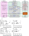
Figure 1
Figure 1
(A) Schematic of steroidogenesis in maternal–fetal compartments during pregnancy. Maternal cholesterol is transported to the placenta, where it serves as a precursor for the production of progesterone and its metabolites. Maternal glucocorticoids can be transported to the placenta and across to the fetal circulation. The fetus makes its own cholesterol from acetate and converts it to DHEAS via several steps, as shown. DHEAS is transported to the fetal liver and from there to the placenta, where DHEAS is converted to testosterone and ultimately to estrogens (estrone (E1), estradiol (E2), and estriol (E3)), which are subsequently transported to the maternal circulation from the placenta. Abbreviations: LDL-cholesterol: low-density lipid cholesterol; CYP11A1: cytochrome P450 family 11 subfamily A member 1; CYP17: cytochrome P450 17; CYP3A7: cytochrome P450 family 3 subfamily A member 7; Δ4A: delta 4 androstenedione; DHEA: dehydroepiandrosterone, DHEAS: dehydroepiandrosterone sulfate; HSD3β1: hydroxysteroid dehydrogenase 3-beta isomerase 1; HSD17β1: hydroxysteroid 17-beta dehydrogenase 1; HSD11β1: hydroxysteroid 11-beta dehydrogenase 1; HSD11β2: hydroxysteroid 11-beta dehydrogenase 2; PAPS: 3′-phospho-5′-adenylyl sulfate; POR: P450 oxidoreductase; STAR: steroidogenic acute regulatory protein; SULT2A1: sulfotransferase family 2A member 1. (B) Maternal stress associates with her health outcomes but does not affect the baby’s birth weight. Linear regression analysis revealed that the maternal perceived stress score (PSS) correlates positively with her sociodemographic score (r = 0.73, p = 0.0008) and body mass index (BMI) (r = 0.48, p = 0.05) but not the infant’s birth weight. The mother’s sociodemographic score positively correlates with her BMI (r = 0.59, p = 0.014). N = 17.
Figure 2
Figure 2
Maternal–fetal glucocorticoid metabolite levels and correlation with birth outcomes and the PSS. (A) Maternal–fetal cortisol levels did not differ significantly, but cord cortisone levels were significantly higher than maternal levels (p = 0.004). (B) Individual cord cortisone levels were higher than cortisol levels, whereas maternal cortisone levels were lower than cortisol levels in all individuals except one. Maternal plasma cortisol correlated positively with maternal plasma cortisone (r = 1.00, p = 0.017). (C) Fetal cortisol or cortisone and (D) maternal cortisol or cortisone did not correlate with the PSS. N = 6. (E) Fetal cortisone levels are maintained high as placental cortisone and progesterone inhibit the activity of HSD11β1; individual placental cortisone levels were higher than placental cortisol levels (p = 0.005) and progesterone (P4) levels were ~13-fold higher than cortisone levels (p < 0.0001). N = 17. Mann–Whitney U test and Spearman’s correlation analysis.
Figure 3
Figure 3
Progesterone and its metabolites’ levels in maternal and fetal samples. (A) Graphic illustration of the relationship between progesterone and its metabolites, dihydroprogesterone (DHP) and allopregnanolone (ALLO). Progesterone is converted to DHP by the action of the enzyme 5α-reductase. DHP is converted to the second metabolite ALLO by 3α-HSD. (B) Cord progesterone levels were ~3-fold higher than maternal plasma progesterone levels (p = 0.041; n = 6). Placental progesterone levels were several-fold greater than maternal hair progesterone levels (p < 0.0001). DHP levels were below the level of detection (LOD) in both maternal and fetal samples, whereas ALLO was only detected in placental tissues (n = 17). Mann–Whitney U test and Spearman’s correlation analysis.
Figure 4
Figure 4
Maternal–fetal progesterone and its metabolites and correlation with birth outcomes. (AE) Maternal and fetal progesterone (n = 6) or placental ALLO (n = 17) levels did not significantly correlate with infant birth weight or gestational age. Spearman’s correlation analysis.
Figure 5
Figure 5
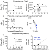
Maternal not fetal progesterone positively correlates with the PSS. (A) Cord or placental progesterone levels did not correlate with the maternal PSS. (B) Placental ALLO levels also did not correlate with the PSS. Placental progesterone levels were ~29-fold higher than placental ALLO levels, and ALLO levels were lower than P4 in each participant. (C) While maternal plasma progesterone levels did not correlate with the PSS, hair P4 levels displayed a significant positive correlation with the PSS (r = 0.964, p = 0.003). N = 7. Mann–Whitney U test and Spearman’s correlation analysis.
Figure 6
Figure 6
Maternal plasma DHEAS levels negatively correlate with the PSS. (A) Cord DHEAS levels were ~7-fold higher than maternal plasma DHEAS levels (p = 0.008). (B) Cord DHEAS levels did not correlate significantly with the PSS or gestational age. (C) Maternal plasma DHEAS levels correlated negatively with the PSS (p = 0.017) but not with gestational age. Mann–Whitney U test and Spearman’s correlation analysis.
Figure 7
Figure 7
Maternal–fetal estrogen metabolites and correlation with birth outcomes and the PSS. (A) Cord estriol levels were ~20-fold higher than estradiol levels and increased in each individual. In the placenta, estrone and estriol levels were higher than estradiol levels, which were lowest in each participant. (B) While neither cord E2 nor E3 levels correlated with gestational age, E2 levels were significantly negatively correlated with the PSS (r = −0.94, p = 0.017). (C) Placental estrogens did not correlate with gestational age or the PSS. (D) Plasma E2 levels showed a strong negative correlation with gestational age, with a trend toward significance (r = −0.90, p = 0.08). Mann–Whitney U test and Spearman’s correlation analysis.

Similar articles

Cited by

References

    1. Ding X.X., Wu Y.L., Xu S.J., Zhu R.P., Jia X.M., Zhang S.F., Huang K., Zhu P., Hao J.H., Tao F.B. Maternal anxiety during pregnancy and adverse birth outcomes: A systematic review and meta-analysis of prospective cohort studies. J. Affect. Disord. 2014;159:103–110. doi: 10.1016/j.jad.2014.02.027. - DOI - PubMed
    1. Khashan A.S., McNamee R., Abel K.M., Mortensen P.B., Kenny L.C., Pedersen M.G., Webb R.T., Baker P.N. Rates of preterm birth following antenatal maternal exposure to severe life events: A population-based cohort study. Hum. Reprod. 2009;24:429–437. doi: 10.1093/humrep/den418. - DOI - PubMed
    1. Khashan A.S., McNamee R., Abel K.M., Pedersen M.G., Webb R.T., Kenny L.C., Mortensen P.B., Baker P.N. Reduced infant birthweight consequent upon maternal exposure to severe life events. Psychosom. Med. 2008;70:688–694. doi: 10.1097/PSY.0b013e318177940d. - DOI - PubMed
    1. Burton G.J., Jauniaux E. What is the placenta? Am. J. Obstet. Gynecol. 2015;213:S6.e1–S6.e4. doi: 10.1016/j.ajog.2015.07.050. - DOI - PubMed
    1. Maltepe E., Fisher S.J. Placenta: The Forgotten Organ. Annu. Rev. Cell Dev. Biol. 2015;31:523–552. doi: 10.1146/annurev-cellbio-100814-125620. - DOI - PubMed

Publication types